Liu Lu, Cai Wenjun, Zhou Chenyang, Tian Hongyan, Wu Beibei, Zhang Jing, Yue Guanghui, Hao Yi
Department of Ultrasound Medicine, South China Hospital, Medical School, Shenzhen University, Shenzhen, P. R. China.
Department of Ultrasound, Shenzhen University General Hospital, Medical School, Shenzhen University, Shenzhen, P. R. China.
Front Med (Lausanne). 2024 Mar 8;11:1362588. doi: 10.3389/fmed.2024.1362588. eCollection 2024.
Accurately differentiating between ovarian endometrioma and ovarian dermoid cyst is of clinical significance. However, the ultrasound appearance of these two diseases is variable, occasionally causing confusion and overlap with each other. This study aimed to develop a diagnostic classification model based on ultrasound radiomics to intelligently distinguish and diagnose the two diseases.
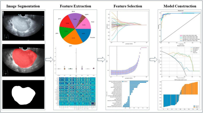
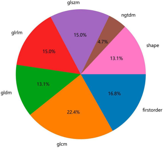
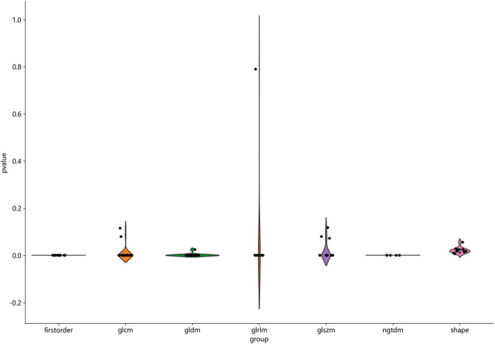
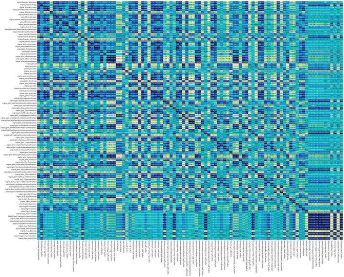
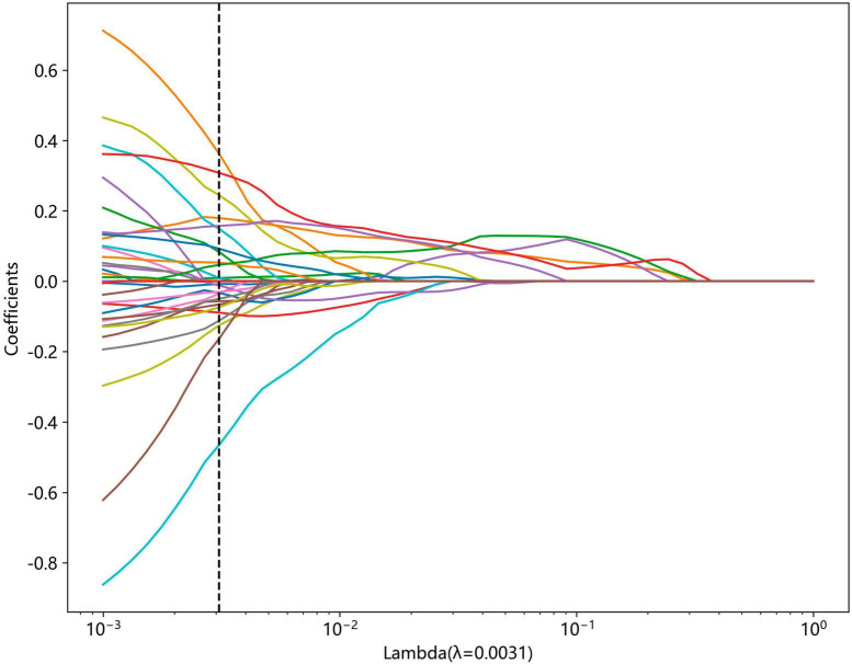
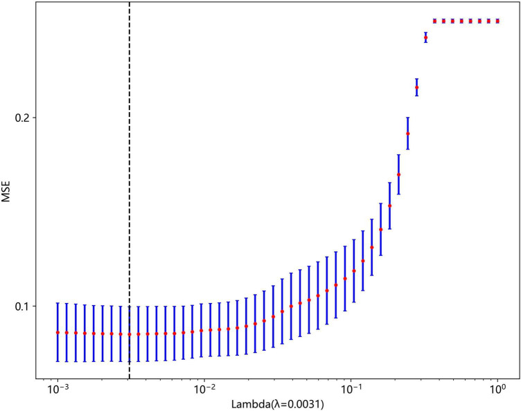
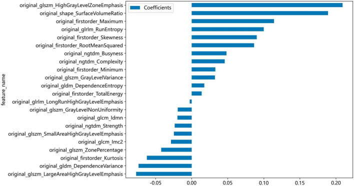
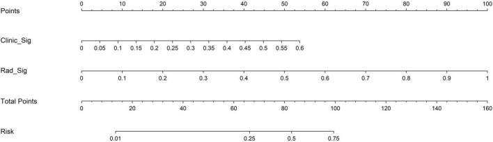
We collected ovarian ultrasound images from participants diagnosed as patients with ovarian endometrioma or ovarian dermoid cyst. Feature extraction and selection were performed using the Mann-Whitney -test, Spearman correlation analysis, and the least absolute shrinkage and selection operator (LASSO) regression. We then input the final features into the machine learning classifiers for model construction. A nomogram was established by combining the radiomic signature and clinical signature.
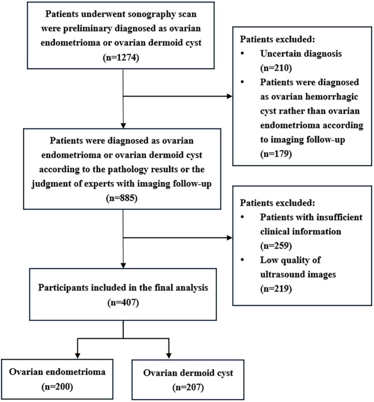
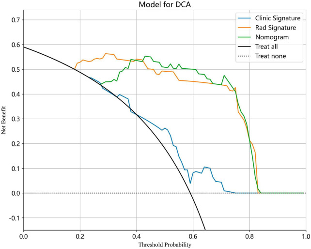
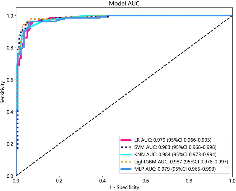
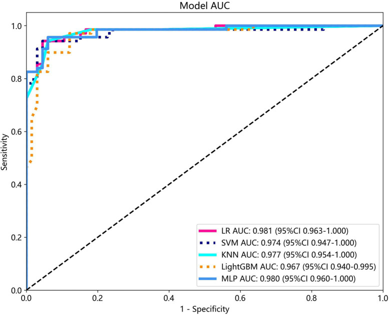
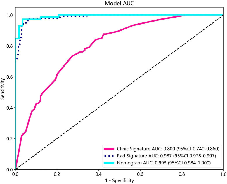
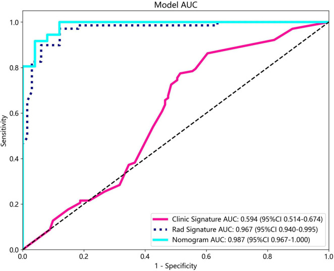
A total of 407 participants with 407 lesions were included and categorized into the ovarian endometriomas group ( = 200) and the dermoid cyst group ( = 207). In the test cohort, Logistic Regression (LR) achieved the highest area under curve (AUC) value (0.981, 95% CI: 0.963-1.000), the highest accuracy (94.8%), and the highest sensitivity (95.5%), while LightGBM achieved the highest specificity (97.1%). A nomogram incorporating both clinical features and radiomic features achieved the highest level of performance (AUC: 0.987, 95% CI: 0.967-1.000, accuracy: 95.1%, sensitivity: 88.0%, specificity: 100.0%, PPV: 100.0%, NPV: 88.0%, precision: 93.6%). No statistical difference in diagnostic performance was observed between the radiomic model and the nomogram ( > 0.05). The diagnostic indexes of radiomic model were comparable to that of senior radiologists and superior to that of junior radiologist. The diagnostic performance of junior radiologists significantly improved with the assistance of the model.
This ultrasound radiomics-based model demonstrated superior diagnostic performance compared to those of junior radiologists and comparable diagnostic performance to those of senior radiologists, and it has the potential to enhance the diagnostic performance of junior radiologists.
准确鉴别卵巢子宫内膜异位囊肿和卵巢皮样囊肿具有临床意义。然而,这两种疾病的超声表现具有多样性,偶尔会导致相互混淆和重叠。本研究旨在基于超声影像组学开发一种诊断分类模型,以智能区分和诊断这两种疾病。
我们收集了被诊断为卵巢子宫内膜异位囊肿或卵巢皮样囊肿患者的卵巢超声图像。使用曼-惠特尼检验、斯皮尔曼相关性分析和最小绝对收缩和选择算子(LASSO)回归进行特征提取和选择。然后将最终特征输入机器学习分类器进行模型构建。通过结合影像组学特征和临床特征建立列线图。
共纳入407例有407个病灶的参与者,分为卵巢子宫内膜异位囊肿组(n = 200)和皮样囊肿组(n = 207)。在测试队列中,逻辑回归(LR)的曲线下面积(AUC)值最高(0.981,95%CI:0.963 - 1.000)、准确率最高(94.8%)和灵敏度最高(95.5%),而LightGBM的特异性最高(97.1%)。结合临床特征和影像组学特征的列线图表现最佳(AUC:0.987,95%CI:0.967 - 1.000,准确率:95.1%,灵敏度:88.0%,特异性:100.0%,阳性预测值:100.0%,阴性预测值:88.0%,精确率:93.6%)。影像组学模型和列线图在诊断性能上无统计学差异(P > 0.05)。影像组学模型的诊断指标与资深放射科医生相当,优于初级放射科医生。在模型的辅助下,初级放射科医生的诊断性能显著提高。
与初级放射科医生相比,这种基于超声影像组学的模型具有卓越的诊断性能,与资深放射科医生相当,并且有可能提高初级放射科医生的诊断性能。