Department of Neurology and the UAB Epilepsy Center, Heersink School of Medicine, University of Alabama at Birmingham, Birmingham, Alabama, USA.
LivaNova Inc, Houston, Texas, USA.
Ann Clin Transl Neurol. 2024 May;11(5):1135-1147. doi: 10.1002/acn3.52029. Epub 2024 Mar 26.
In parallel to standard vagus nerve stimulation (VNS), microburst stimulation delivery has been developed. We evaluated the fMRI-related signal changes associated with standard and optimized microburst stimulation in a proof-of-concept study (NCT03446664).
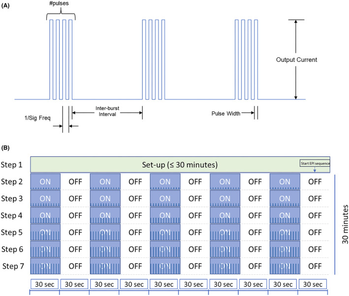
Twenty-nine drug-resistant epilepsy patients were prospectively implanted with VNS. Three 3T fMRI scans were collected 2 weeks postimplantation. The maximum tolerated VNS intensity was determined prior to each scan starting at 0.125 mA with 0.125 mA increments. FMRI scans were block-design with alternating 30 sec stimulation [ON] and 30 sec no stimulation [OFF]: Scan 1 utilized standard VNS and Scan 3 optimized microburst parameters to determine target settings. Semi-automated on-site fMRI data processing utilized ON-OFF block modeling to determine VNS-related fMRI activation per stimulation setting. Anatomical thalamic mask was used to derive highest mean thalamic t-value for determination of microburst stimulation parameters. Paired t-tests corrected at P < 0.05 examined differences in fMRI responses to each stimulation type.
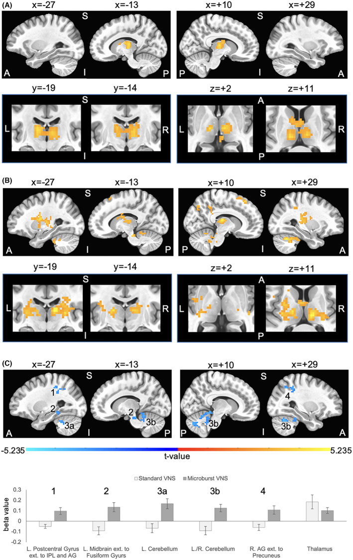
Standard and microburst stimulation intensities at Scans 1 and 3 were similar (P = 0.16). Thalamic fMRI responses were obtained in 28 participants (19 with focal; 9 with generalized seizures). Group activation maps showed standard VNS elicited thalamic activation while optimized microburst VNS showed widespread activation patterns including thalamus. Comparison of stimulation types revealed significantly greater cerebellar, midbrain, and parietal fMRI signal changes in microburst compared to standard VNS. These differences were not associated with seizure responses.
While standard and optimized microburst VNS elicited thalamic activation, microburst also engaged other brain regions. Relationship between these fMRI activation patterns and clinical response warrants further investigation.
The study was registered with clinicaltrials.gov (NCT03446664).
与标准迷走神经刺激 (VNS) 并行,已经开发出微爆发刺激传递。我们在一项概念验证研究中评估了与标准和优化微爆发刺激相关的 fMRI 相关信号变化(NCT03446664)。
29 名耐药性癫痫患者前瞻性植入 VNS。在植入后 2 周收集了 3 次 3T fMRI 扫描。在每次扫描开始时,以 0.125mA 的增量确定最大耐受 VNS 强度,起始强度为 0.125mA。FMRI 扫描采用交替 30 秒刺激 [ON] 和 30 秒无刺激 [OFF] 的块设计:扫描 1 使用标准 VNS,扫描 3 使用优化的微爆发参数确定目标设置。半自动化现场 fMRI 数据处理利用 ON-OFF 块建模来确定每种刺激设置的 VNS 相关 fMRI 激活。使用解剖丘脑掩模来确定微爆发刺激参数的最高平均丘脑 t 值。配对 t 检验(校正 P < 0.05)检查了两种刺激类型的 fMRI 反应差异。
扫描 1 和 3 的标准和微爆发刺激强度相似(P = 0.16)。在 28 名参与者中获得了丘脑 fMRI 反应(19 名有局灶性;9 名有全身性发作)。组激活图显示标准 VNS 引起了丘脑激活,而优化的微爆发 VNS 显示了广泛的激活模式,包括丘脑。刺激类型的比较显示,与标准 VNS 相比,微爆发 VNS 引起了小脑、中脑和顶叶 fMRI 信号的显著变化。这些差异与癫痫发作反应无关。
虽然标准和优化的微爆发 VNS 引起了丘脑激活,但微爆发还涉及其他大脑区域。这些 fMRI 激活模式与临床反应之间的关系需要进一步研究。
该研究在 clinicaltrials.gov 上注册(NCT03446664)。