Paczkowska-Walendowska Magdalena, Koumentakou Ioanna, Lazaridou Maria, Bikiaris Dimitrios, Miklaszewski Andrzej, Plech Tomasz, Cielecka-Piontek Judyta
Department of Pharmacognosy and Biomaterials, Poznan University of Medical Sciences, 60-806 Poznan, Poland.
Laboratory of Polymer Chemistry and Technology, Department of Chemistry, Aristotle University of Thessaloniki, 541 24 Thessaloniki, Greece.
Pharmaceutics. 2024 Mar 4;16(3):359. doi: 10.3390/pharmaceutics16030359.
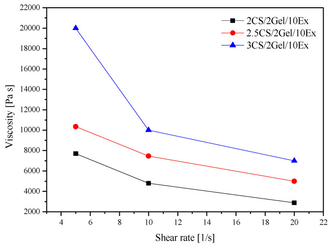
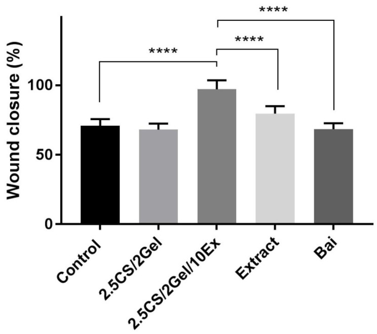
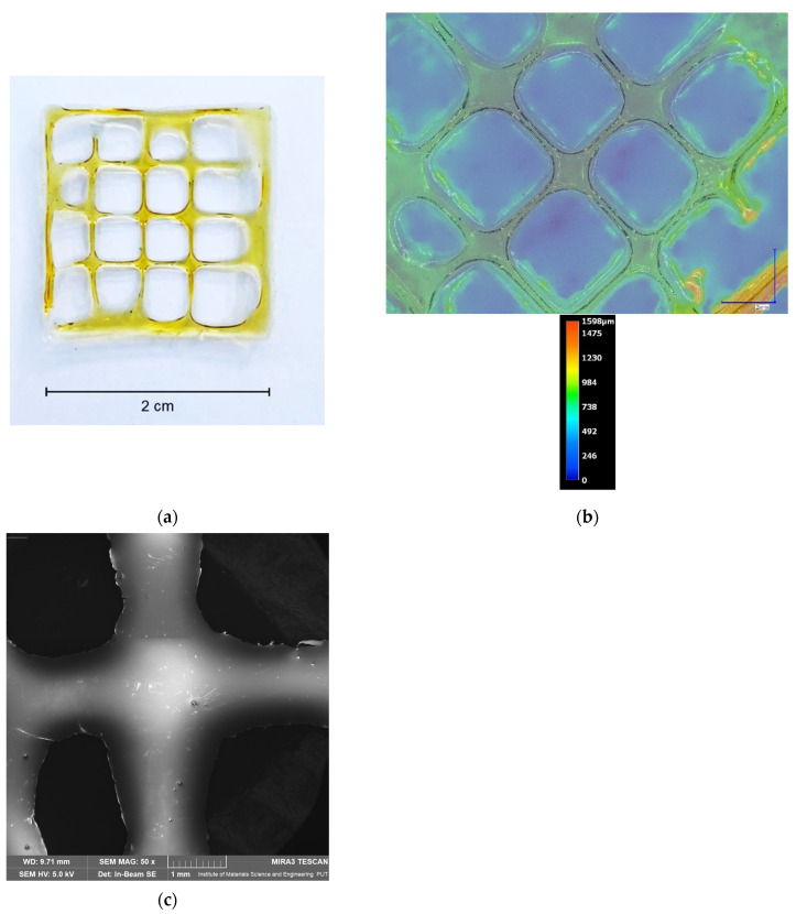
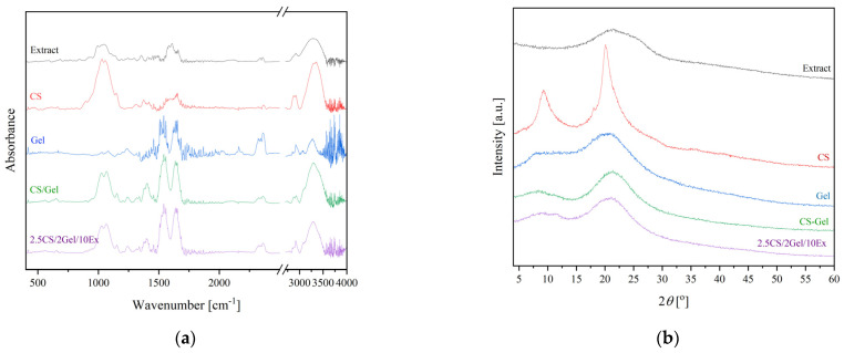
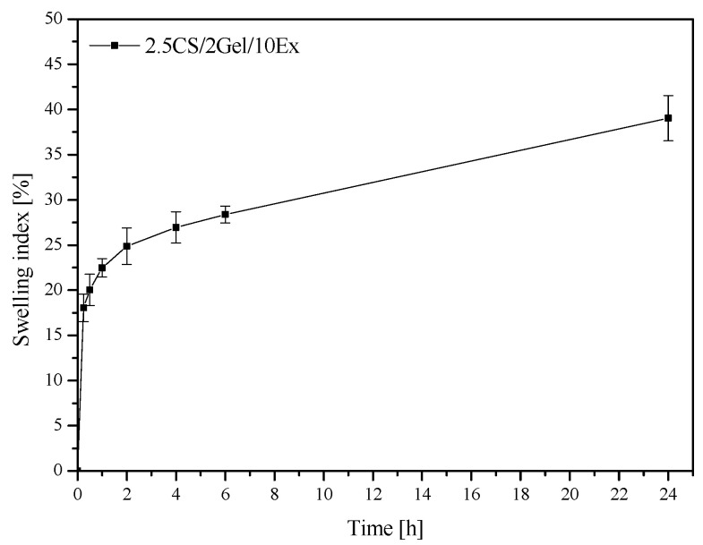
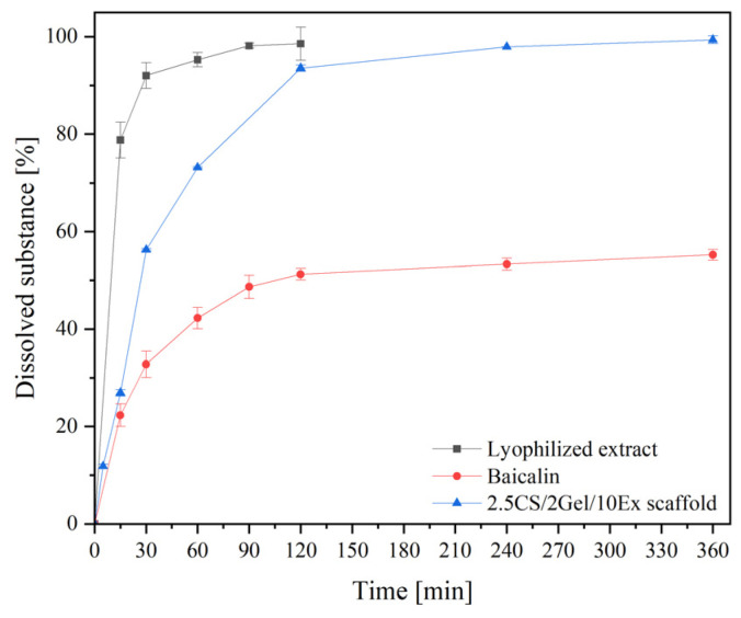
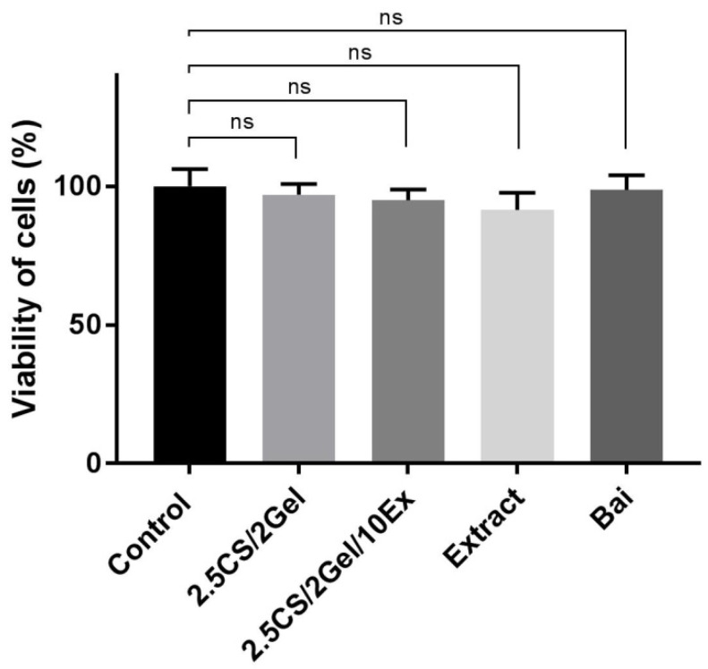
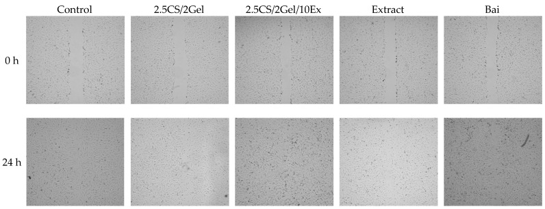
The plant material , which is rich in flavones (baicalin), possesses antibacterial, antifungal, antioxidant, and anti-inflammatory properties. This work aimed to develop a 3D-printed chitosan-based hydrogel rich in extract as an innovative approach for the personalized treatment of periodontal diseases. Chitosan-based hydrogels were prepared, and the printability of the prepared hydrogels was determined. The hydrogel with 2.5% / of high molecular-weight chitosan (CS), 2% / gelatin (Gel), and 10% / of extract (Ex) presented the best printability, producing smooth and uniform scaffolds. It was proved that the CS/Gel/Ex hydrogel was stabilized by hydrogen bonds and remained in amorphous dispersion in the 3D-printed structures (confirmed by ATR-FTIR and XRPD). Due to the amorphization of the active substance, a significant increase in the release of baicalin in vitro was observed. It was demonstrated that there was an initial burst release and a continuous release profile ( = 3). Higuchi kinetic was the most likely baicalin release kinetic. The second fit, the Korsmeyer-Peppas kinetics model, showed coupled diffusion of the active ingredient in the hydrated matrix and polymer relaxation regulated release, with n values ranging from 0.45 to 0.89. The anti-inflammatory properties of 3D-printed scaffolds were assessed as the ability to inhibit the activity of the hyaluronidase enzyme. Activity was assessed as IC = 63.57 ± 4.98 mg hydrogel/mL ( = 6). Cytotoxicity tests demonstrated the biocompatibility of the material. After 24 h of exposure to the 2.5CS/2Gel/10Ex scaffold, fibroblasts migrated toward the scratch, closed the "wound" by 97.1%, and significantly accelerated the wound healing process. The results render the 3D-printed CS/Gel/extract scaffolds as potential candidates for treating periodontal diseases.
这种富含黄酮类化合物(黄芩苷)的植物材料具有抗菌、抗真菌、抗氧化和抗炎特性。这项工作旨在开发一种富含提取物的3D打印壳聚糖基水凝胶,作为一种创新方法用于牙周疾病的个性化治疗。制备了壳聚糖基水凝胶,并测定了所制备水凝胶的可打印性。含有2.5%/的高分子量壳聚糖(CS)、2%/明胶(Gel)和10%/提取物(Ex)的水凝胶表现出最佳的可打印性,能制造出光滑且均匀的支架。事实证明,CS/Gel/Ex水凝胶通过氢键稳定,并在3D打印结构中保持无定形分散状态(通过ATR-FTIR和XRPD证实)。由于活性物质的非晶化,观察到黄芩苷在体外的释放显著增加。结果表明存在初始突释和持续释放曲线(n = 3)。Higuchi动力学最有可能是黄芩苷的释放动力学。二次拟合,即Korsmeyer-Peppas动力学模型,显示活性成分在水合基质中的耦合扩散以及聚合物松弛调节释放,n值范围为0.45至0.89。3D打印支架的抗炎特性通过抑制透明质酸酶活性的能力来评估。活性评估为IC = 63.57 ± 4.98 mg水凝胶/mL(n = 6)。细胞毒性测试证明了该材料的生物相容性。在暴露于2.5CS/2Gel/10Ex支架24小时后,成纤维细胞向划痕处迁移,将“伤口”闭合了97.1%,并显著加速了伤口愈合过程。这些结果使3D打印的CS/Gel/提取物支架成为治疗牙周疾病的潜在候选材料。