Department of Radiation and Medical Oncology, Zhongnan Hospital of Wuhan University, Wuhan, Hubei, 430071, China.
Hubei Key Laboratory of Tumor Biological Behaviors, Zhongnan Hospital, Wuhan University, Wuhan, China.
Mol Cancer. 2024 Apr 4;23(1):70. doi: 10.1186/s12943-024-01985-1.
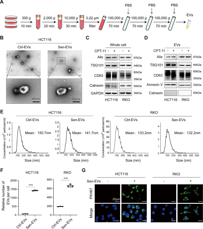
Cellular senescence frequently occurs during anti-cancer treatment, and persistent senescent tumor cells (STCs) unfavorably promote tumor progression through paracrine secretion of the senescence-associated secretory phenotype (SASP). Extracellular vesicles (EVs) have recently emerged as a novel component of the SASP and primarily mediate the tumor-promoting effect of the SASP. Of note, the potential effect of EVs released from STCs on tumor progression remains largely unknown.
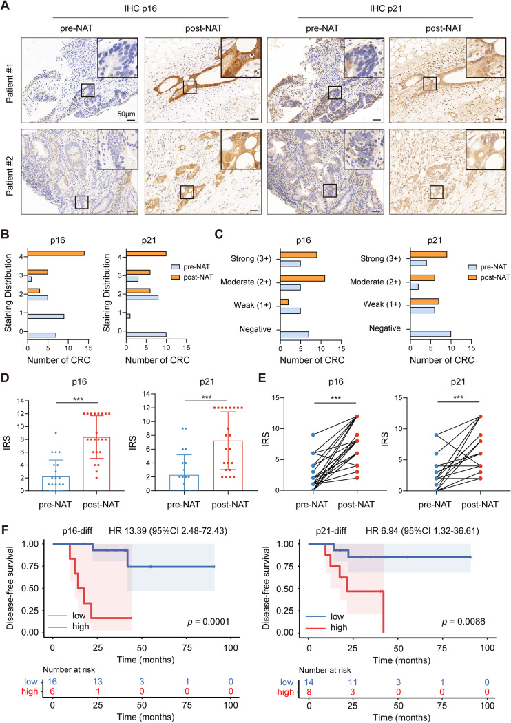
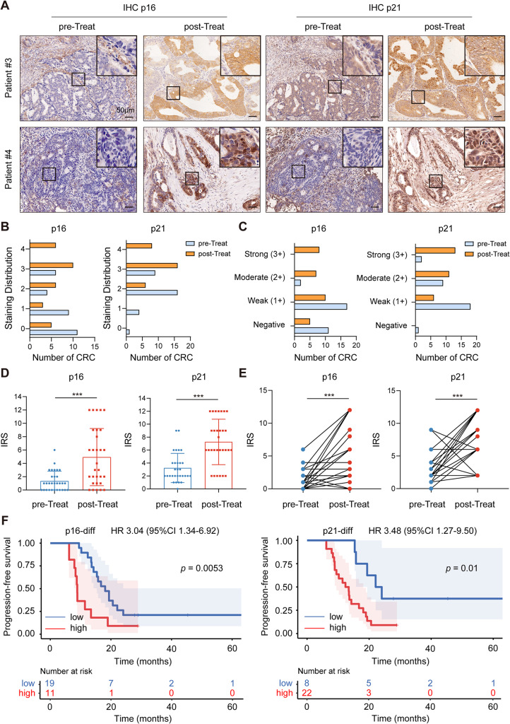
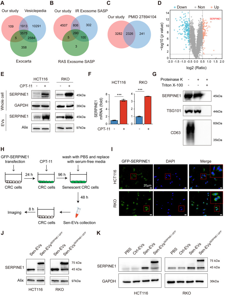
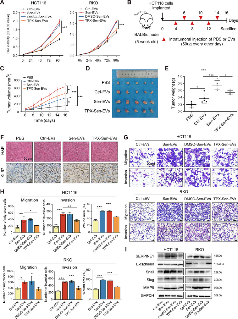
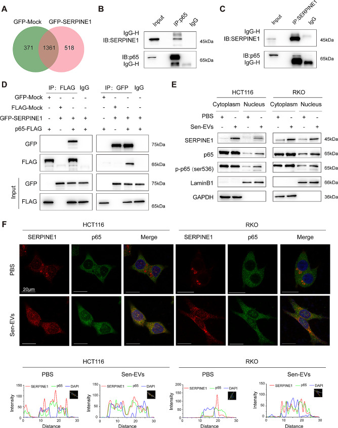
We collected tumor tissues from two cohorts of colorectal cancer (CRC) patients to examine the expression of p16, p21, and SERPINE1 before and after anti-cancer treatment. Cohort 1 included 22 patients with locally advanced rectal cancer (LARC) who received neoadjuvant therapy before surgical resection. Cohort 2 included 30 patients with metastatic CRC (mCRC) who received first-line irinotecan-contained treatment. CCK-8, transwell, wound-healing assay, and tumor xenograft experiments were carried out to determine the impacts of EVs released from STCs on CRC progression in vitro and in vivo. Quantitative proteomic analysis was applied to identify protein cargo inside EVs secreted from STCs. Immunoprecipitation and mass spectrometer identification were utilized to explore the binding partners of SERPINE1. The interaction of SERPINE1 with p65 was verified by co-immunoprecipitation, and their co-localization was confirmed by immunofluorescence.
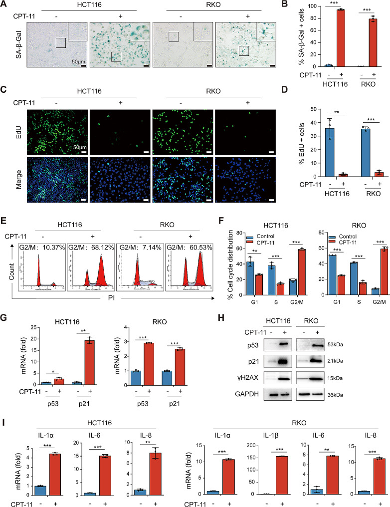
Chemotherapeutic agents and irradiation could potently induce senescence in CRC cells in vitro and in human CRC tissues. The more significant elevation of p16 and p21 expression in patients after anti-cancer treatment displayed shorter disease-free survival (DFS) for LARC or progression-free survival (PFS) for mCRC. We observed that compared to non-STCs, STCs released an increased number of EVs enriched in SERPINE1, which further promoted the progression of recipient cancer cells. Targeting SERPINE1 with a specific inhibitor, tiplaxtinin, markedly attenuated the tumor-promoting effect of STCs-derived EVs. Additionally, the patients with greater increment of SERPINE1 expression after anti-cancer treatment had shorter DFS for LARC or PFS for mCRC. Mechanistically, SERPINE1 bound to p65, promoting its nuclear translocation and subsequently activating the NF-κB signaling pathway.
We provide the in vivo evidence of the clinical prognostic implications of therapy-induced senescence. Our results revealed that STCs were responsible for CRC progression by producing large amounts of EVs enriched in SERPINE1. These findings further confirm the crucial role of therapy-induced senescence in tumor progression and offer a potential therapeutic strategy for CRC treatment.
细胞衰老在抗癌治疗中经常发生,持续存在的衰老肿瘤细胞(STC)通过衰老相关分泌表型(SASP)的旁分泌分泌不利地促进肿瘤进展。细胞外囊泡(EVs)最近成为 SASP 的一个新组成部分,主要介导 SASP 的促肿瘤作用。值得注意的是,STC 释放的 EVs 对肿瘤进展的潜在影响在很大程度上尚不清楚。
我们从两批结直肠癌(CRC)患者的肿瘤组织中收集样本,以检查抗癌治疗前后 p16、p21 和 SERPINE1 的表达。队列 1 包括 22 名接受新辅助治疗后手术切除的局部晚期直肠癌(LARC)患者。队列 2 包括 30 名接受一线伊立替康治疗的转移性 CRC(mCRC)患者。CCK-8、Transwell、伤口愈合试验和肿瘤异种移植实验用于确定体外和体内 STC 释放的 EVs 对 CRC 进展的影响。定量蛋白质组学分析用于鉴定 STC 分泌的 EV 中的蛋白质货物。免疫沉淀和质谱鉴定用于探索 SERPINE1 的结合伙伴。通过共免疫沉淀验证了 SERPINE1 与 p65 的相互作用,并通过免疫荧光确认了它们的共定位。
化疗药物和辐射可以在体外和人 CRC 组织中强烈诱导 CRC 细胞衰老。抗癌治疗后患者 p16 和 p21 表达升高更明显,LARC 的无病生存期(DFS)或 mCRC 的无进展生存期(PFS)更短。我们观察到,与非 STC 相比,STC 释放的 EVs 数量增加,其中富含 SERPINE1,进一步促进了受体癌细胞的进展。用特异性抑制剂 tiplaxtinin 靶向 SERPINE1,显著减弱了 STC 衍生 EVs 的促肿瘤作用。此外,抗癌治疗后 SERPINE1 表达增加更大的患者 LARC 的 DFS 或 mCRC 的 PFS 更短。在机制上,SERPINE1 与 p65 结合,促进其核转位,进而激活 NF-κB 信号通路。
我们提供了治疗诱导衰老的临床预后意义的体内证据。我们的结果表明,STC 通过产生富含 SERPINE1 的大量 EVs 导致 CRC 进展。这些发现进一步证实了治疗诱导衰老在肿瘤进展中的关键作用,并为 CRC 治疗提供了一种潜在的治疗策略。