Metabolic Research Laboratories, Wellcome-Medical Research Council Institute of Metabolic Science, University of Cambridge, Cambridge, CB2 0QQ, UK.
MRC Cancer Unit, University of Cambridge, Cambridge Biomedical Campus, Cambridge, CB2 0XZ, UK.
EMBO J. 2024 Jun;43(11):2127-2165. doi: 10.1038/s44318-024-00084-7. Epub 2024 Apr 5.
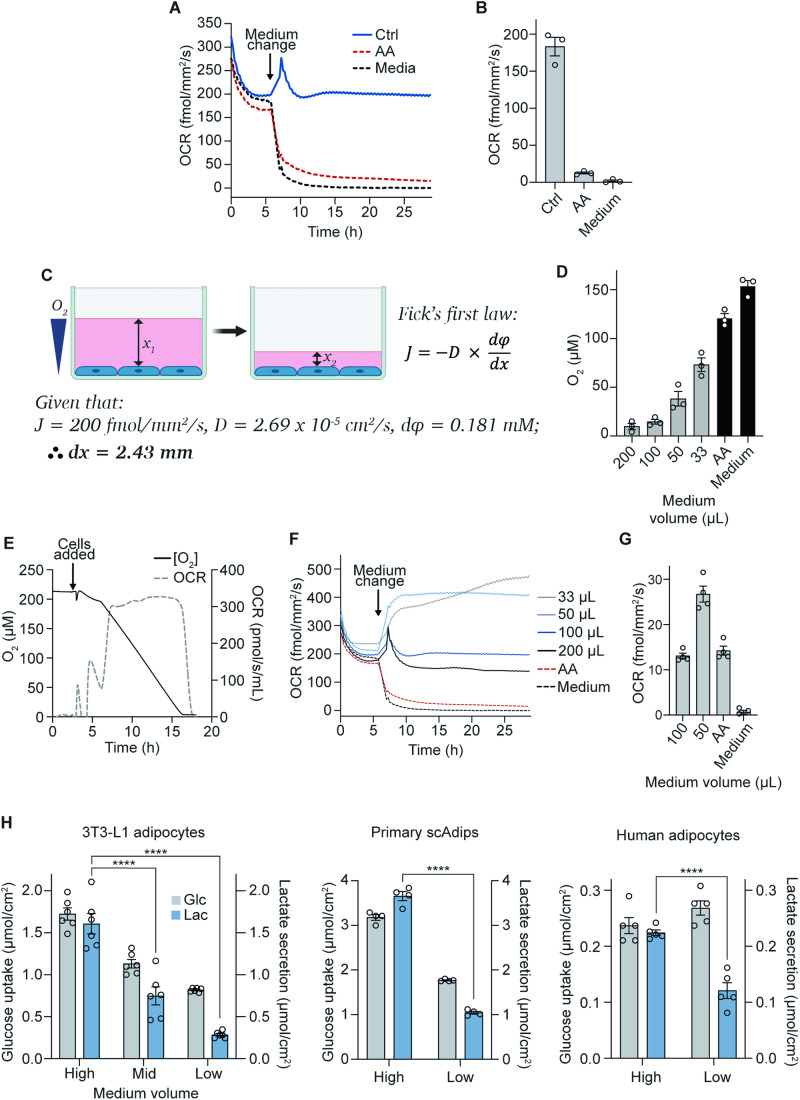
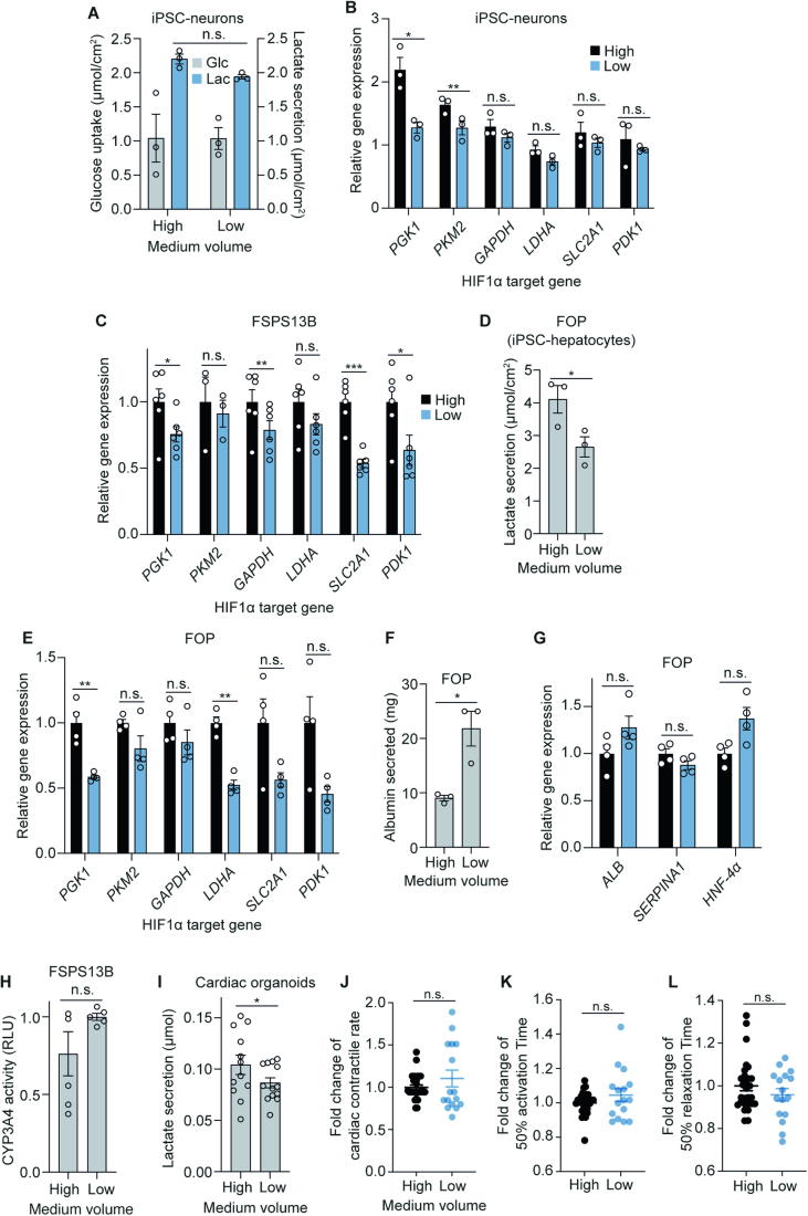
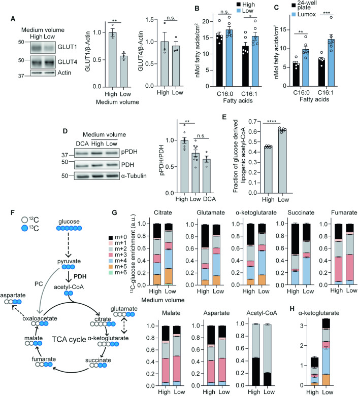
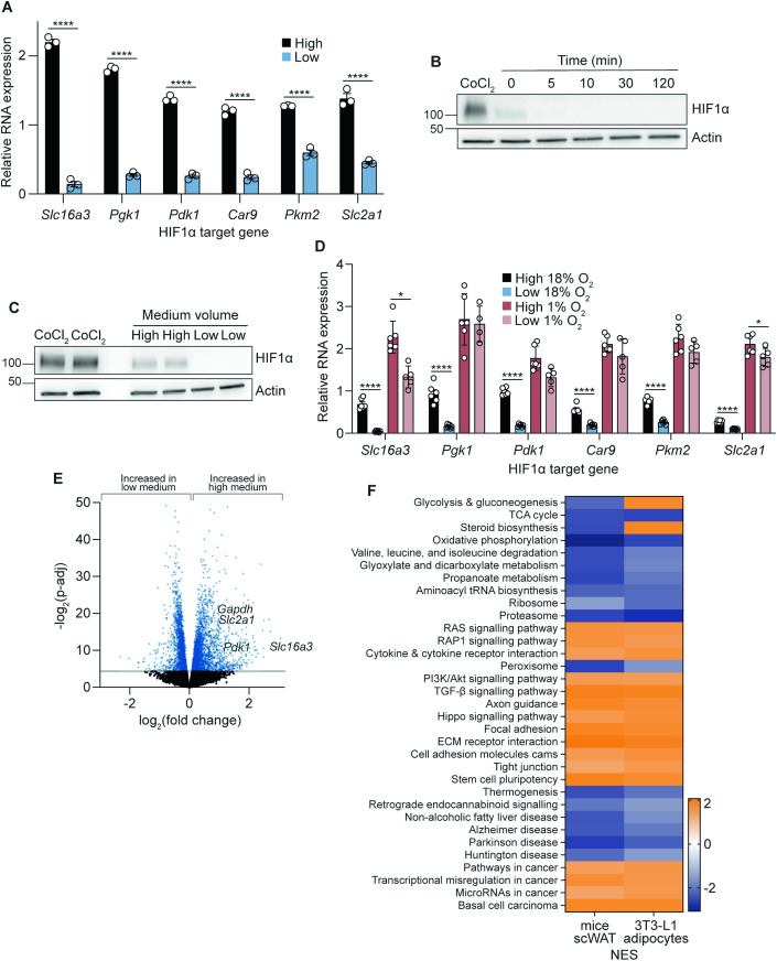
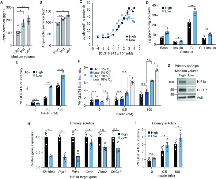
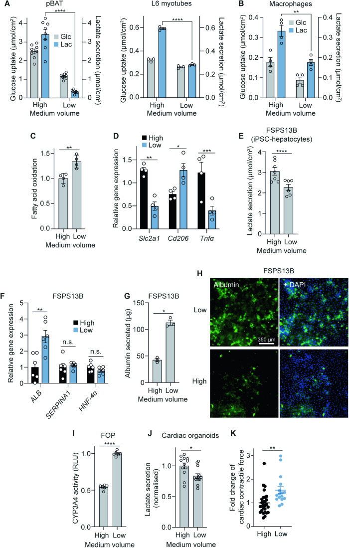
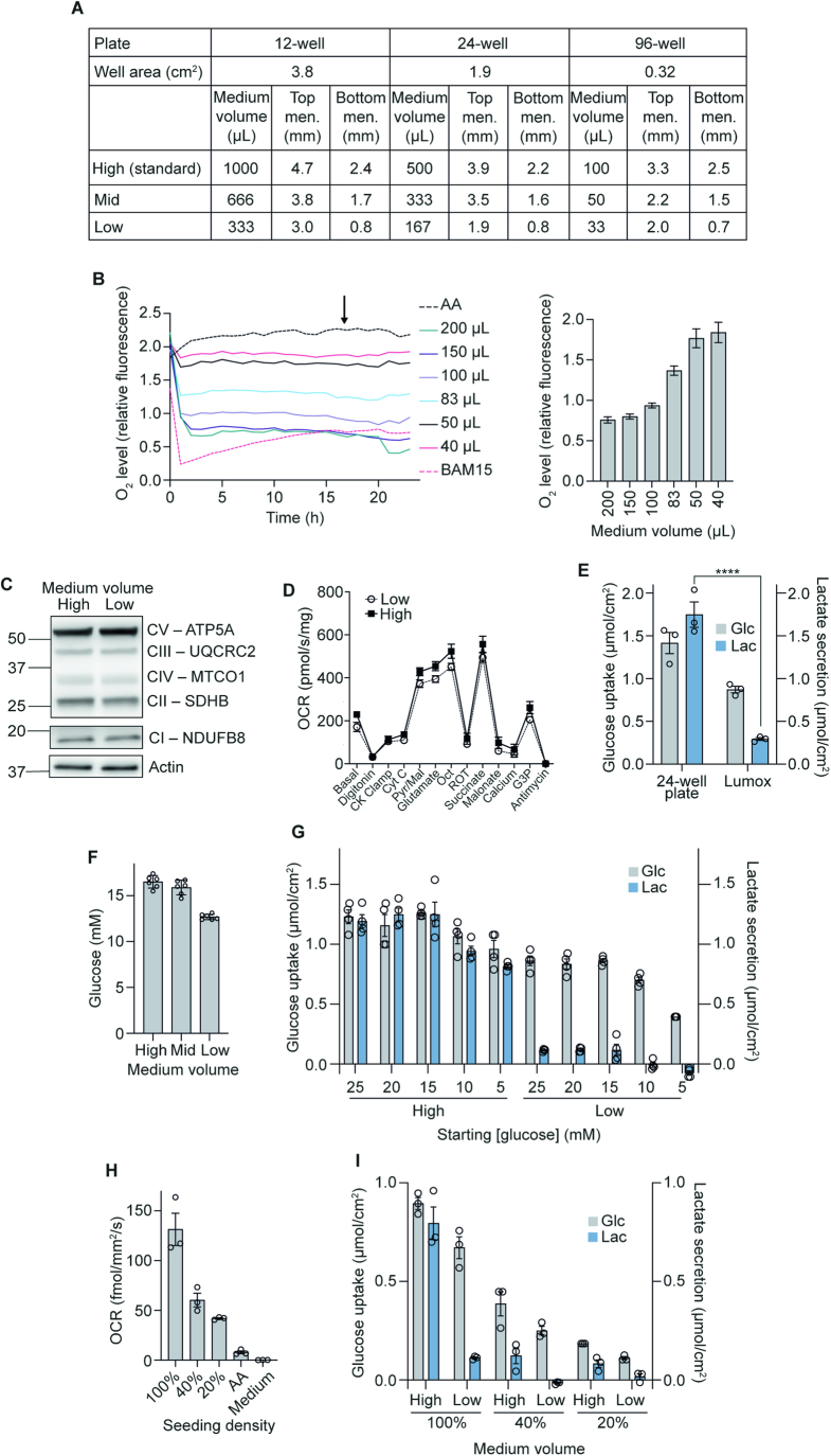
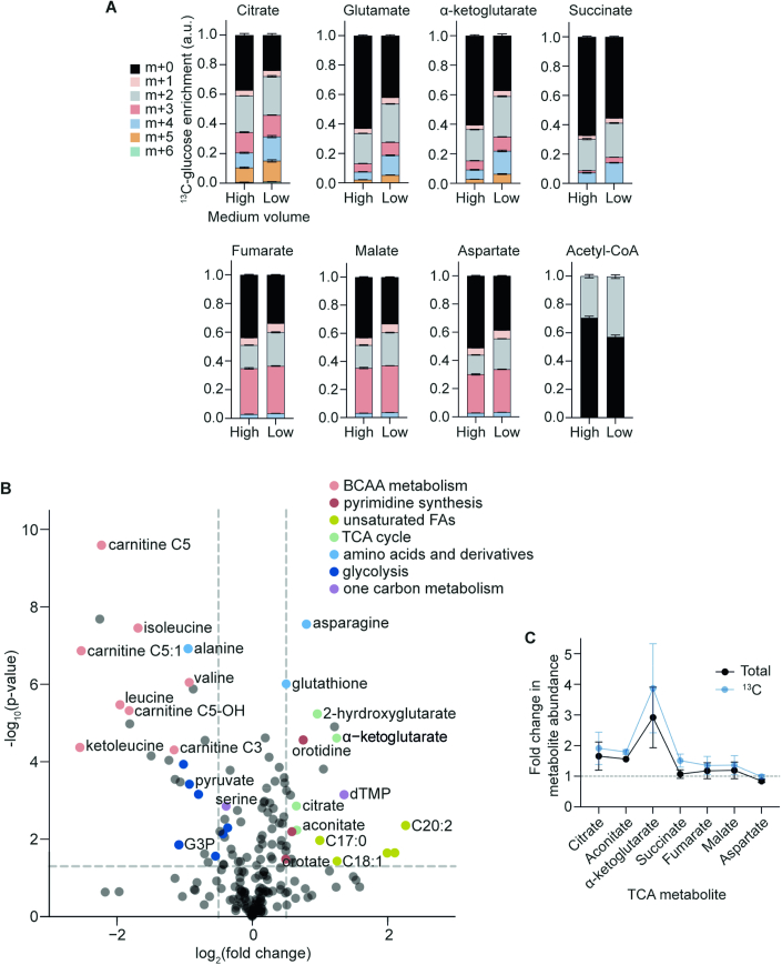
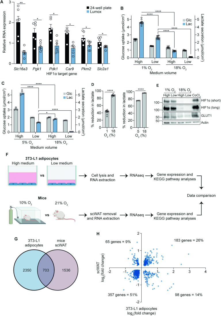
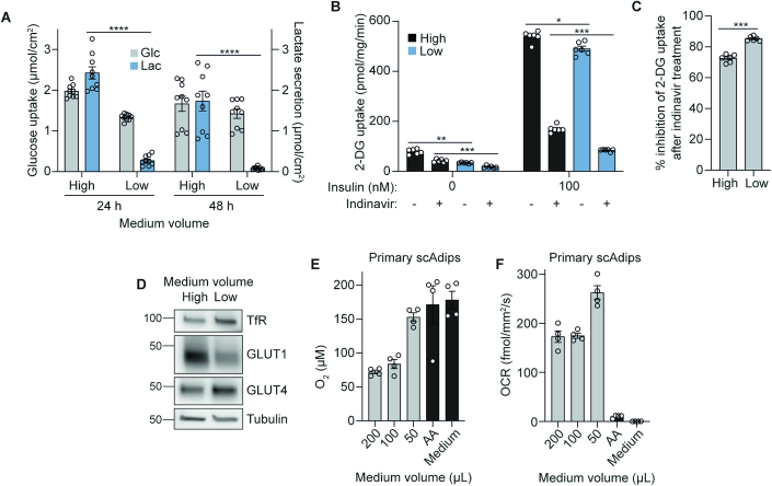
The in vitro oxygen microenvironment profoundly affects the capacity of cell cultures to model physiological and pathophysiological states. Cell culture is often considered to be hyperoxic, but pericellular oxygen levels, which are affected by oxygen diffusivity and consumption, are rarely reported. Here, we provide evidence that several cell types in culture actually experience local hypoxia, with important implications for cell metabolism and function. We focused initially on adipocytes, as adipose tissue hypoxia is frequently observed in obesity and precedes diminished adipocyte function. Under standard conditions, cultured adipocytes are highly glycolytic and exhibit a transcriptional profile indicative of physiological hypoxia. Increasing pericellular oxygen diverted glucose flux toward mitochondria, lowered HIF1α activity, and resulted in widespread transcriptional rewiring. Functionally, adipocytes increased adipokine secretion and sensitivity to insulin and lipolytic stimuli, recapitulating a healthier adipocyte model. The functional benefits of increasing pericellular oxygen were also observed in macrophages, hPSC-derived hepatocytes and cardiac organoids. Our findings demonstrate that oxygen is limiting in many terminally-differentiated cell types, and that considering pericellular oxygen improves the quality, reproducibility and translatability of culture models.
体外氧微环境深刻影响细胞培养物模拟生理和病理状态的能力。细胞培养通常被认为是高氧的,但细胞周围的氧水平受氧扩散和消耗的影响,很少有报道。在这里,我们提供的证据表明,培养中的几种细胞实际上经历局部缺氧,这对细胞代谢和功能有重要影响。我们最初关注脂肪细胞,因为肥胖症中经常观察到脂肪组织缺氧,并且在脂肪细胞功能下降之前就出现了这种情况。在标准条件下,培养的脂肪细胞具有很高的糖酵解活性,并表现出与生理缺氧相关的转录谱。增加细胞周围的氧气会将葡萄糖通量转移到线粒体,降低 HIF1α 的活性,并导致广泛的转录重编程。从功能上讲,脂肪细胞增加了脂肪因子的分泌以及对胰岛素和脂肪分解刺激的敏感性,再现了更健康的脂肪细胞模型。增加细胞周围氧气的功能益处也在巨噬细胞、hPSC 衍生的肝细胞和心脏类器官中观察到。我们的发现表明,许多终末分化的细胞类型中氧是有限的,并且考虑细胞周围氧可以提高培养模型的质量、可重复性和可翻译性。