State Key Laboratory of Oral Diseases &National Clinical Research Center for Oral Diseases & Engineering Research Center of Oral Translational Medicine, Ministry of Education & National Engineering Laboratory for Oral Regenerative Medicine, West China Hospital of Stomatology, Sichuan University, Chengdu, People's Republic of China.
Department of Oral and Maxillofacial Surgery, West China Hospital of Stomatology, Sichuan University, Chengdu, People's Republic of China.
J Extracell Vesicles. 2024 Apr;13(4):e12434. doi: 10.1002/jev2.12434.
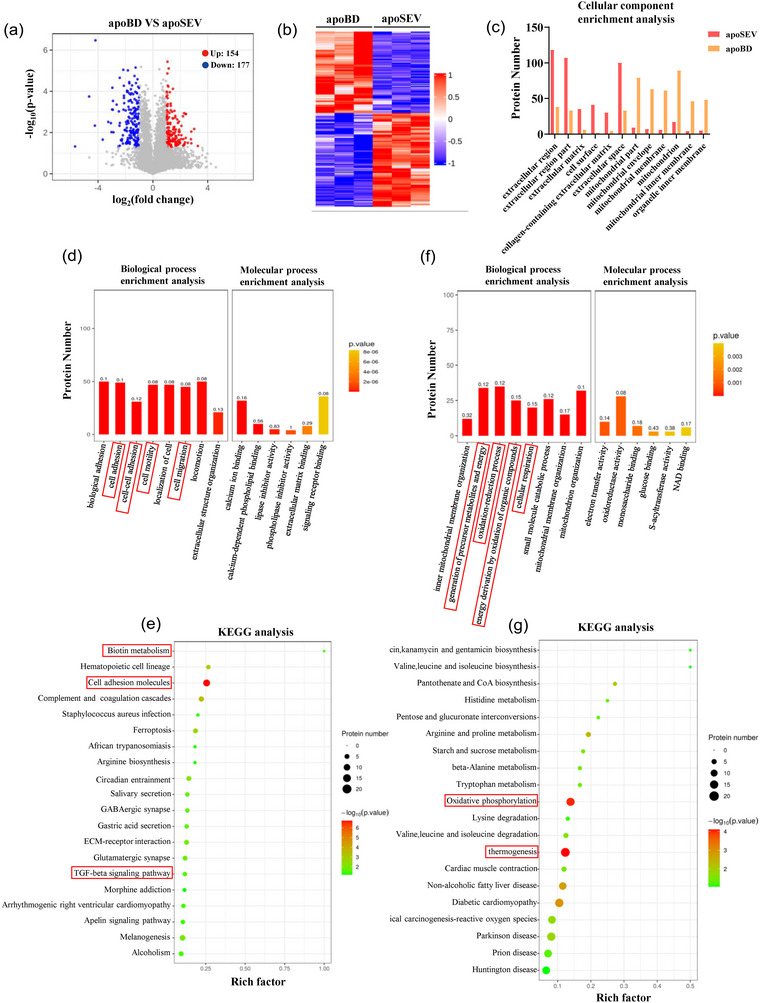
Apoptosis releases numerous apoptotic vesicles that regulate processes such as cell proliferation, immunity, and tissue regeneration and repair. Now, it has also emerged as an attractive candidate for biotherapeutics. However, apoptotic vesicles encompass a diverse range of subtypes, and it remains unclear which specific subtypes play a pivotal role. In this study, we successfully isolated different apoptotic vesicle subtypes based on their sizes and characterized them using NTA and TEM techniques, respectively. We compared the functional variances among the distinct subtypes of apoptotic vesicles in terms of stem cell proliferation, migration, and differentiation, as well as for endothelial cell and macrophage function, effectively identifying subtypes that exhibit discernible functional differences. ApoSEV (with diameter <1000 nm) promoted stem cell proliferation, migration, and multi-potent differentiation, and accelerated skin wound healing of diabetes mouse model, while apoBD (with diameter >1000 nm) played the opposite effect on cell function and tissue regeneration. Lastly, employing protein analysis and gene sequencing techniques, we elucidated the intrinsic mechanisms underlying these differences between different subtypes of apoEVs. Collectively, this study identified that apoptotic vesicle subtypes possessed distinct bio-functions in regulating stem cell function and behaviour and modulating tissue regeneration, which primarily attribute to the distinct profiling of protein and mRNA in different subtypes. This comprehensive analysis of specific subtypes of apoEVs would provide novel insights for potential therapeutic applications in cell biology and tissue regeneration.
细胞凋亡会释放出许多凋亡小体,这些小体调节着细胞增殖、免疫和组织再生和修复等过程。现在,凋亡小体也已经成为生物治疗的一个有吸引力的候选物。然而,凋亡小体包含多种不同的亚型,目前尚不清楚哪种特定的亚型起着关键作用。在这项研究中,我们成功地根据大小分离出不同的凋亡小体亚型,并分别使用 NTA 和 TEM 技术对它们进行了表征。我们比较了不同凋亡小体亚型在干细胞增殖、迁移和分化以及内皮细胞和巨噬细胞功能方面的功能差异,有效地鉴定出具有明显功能差异的亚型。直径<1000nm 的凋亡小体(ApoSEV)促进干细胞增殖、迁移和多能分化,并加速糖尿病小鼠模型的皮肤伤口愈合,而直径>1000nm 的凋亡小体(apoBD)对细胞功能和组织再生则产生相反的影响。最后,我们运用蛋白质分析和基因测序技术,阐明了不同亚型 apoEVs 之间差异的内在机制。总之,这项研究表明,凋亡小体亚型在调节干细胞功能和行为以及调节组织再生方面具有不同的生物功能,这主要归因于不同亚型中蛋白质和 mRNA 的不同特征。对特定亚型 apoEVs 的综合分析将为细胞生物学和组织再生的潜在治疗应用提供新的见解。