McNamara Harold M, Jia Bill Z, Guyer Alison, Parot Vicente J, Dobbs Caleb, Schier Alexander F, Cohen Adam E, Lord Nathan D
Lewis Sigler Institute, Princeton University, Princeton, NJ, USA.
Department of Systems Biology, Blavatnik Institute, Harvard Medical School, Boston, MA, USA.
bioRxiv. 2024 Apr 12:2024.04.11.588875. doi: 10.1101/2024.04.11.588875.
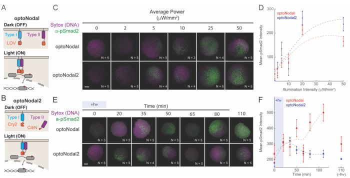
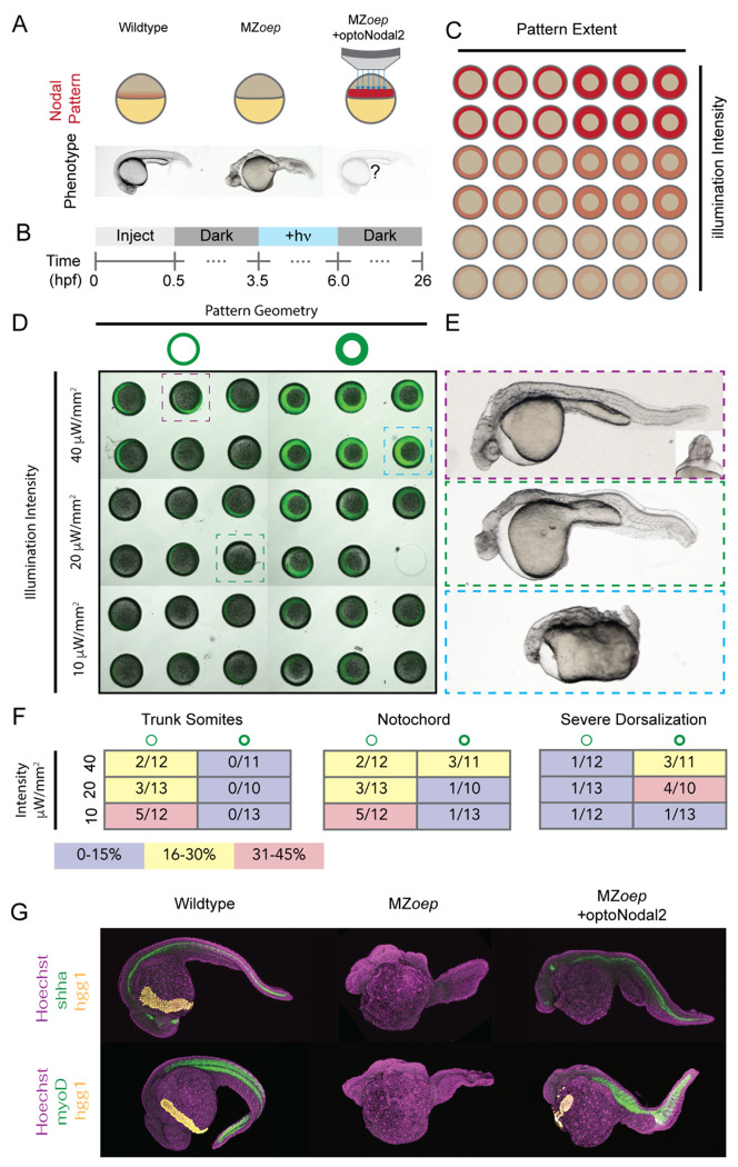
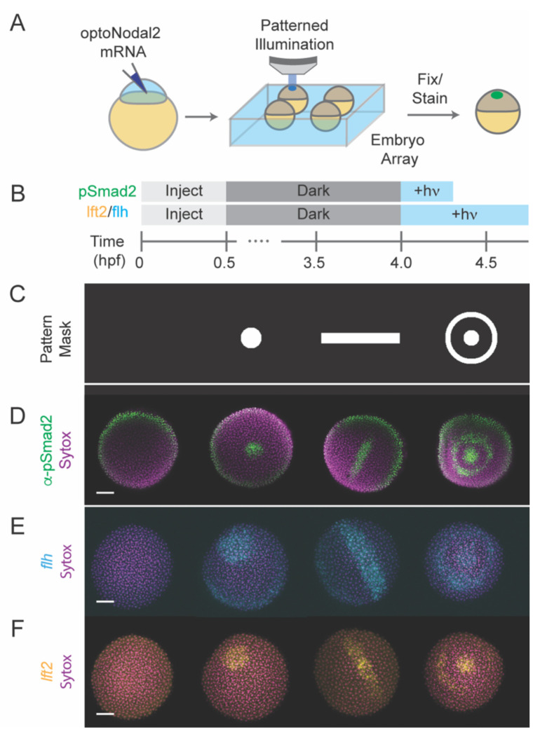
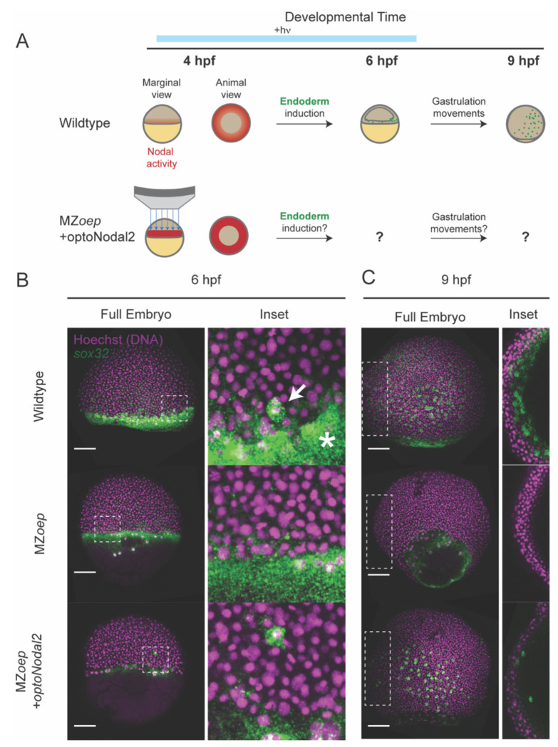
A crucial step in early embryogenesis is the establishment of spatial patterns of signaling activity. Tools to perturb morphogen signals with high resolution in space and time can help reveal how embryonic cells decode these signals to make appropriate fate decisions. Here, we present new optogenetic reagents and an experimental pipeline for creaHng designer Nodal signaling patterns in live zebrafish embryos. Nodal receptors were fused to the light-sensitive heterodimerizing pair Cry2/CIB1N, and the Type II receptor was sequestered to the cytosol. The improved optoNodal2 reagents eliminate dark activity and improve response kinetics, without sacrificing dynamic range. We adapted an ultra-widefield microscopy platform for parallel light patterning in up to 36 embryos and demonstrated precise spatial control over Nodal signaling activity and downstream gene expression. Patterned Nodal activation drove precisely controlled internalization of endodermal precursors. Further, we used patterned illumination to generate synthetic signaling patterns in Nodal signaling mutants, rescuing several characteristic developmental defects. This study establishes an experimental toolkit for systematic exploration of Nodal signaling patterns in live embryos.
早期胚胎发育中的一个关键步骤是信号活性空间模式的建立。能够在空间和时间上高分辨率地扰动形态发生素信号的工具,有助于揭示胚胎细胞如何解码这些信号以做出适当的命运决定。在此,我们展示了新的光遗传学试剂和一个实验流程,用于在活的斑马鱼胚胎中创建定制的Nodal信号模式。将Nodal受体与光敏感异源二聚化对Cry2/CIB1N融合,并将II型受体隔离在细胞质中。改进后的optoNodal2试剂消除了暗活性并改善了反应动力学,同时不牺牲动态范围。我们采用了一个超宽场显微镜平台,用于对多达36个胚胎进行平行光图案化,并展示了对Nodal信号活性和下游基因表达的精确空间控制。图案化的Nodal激活驱动了内胚层前体的精确控制内化。此外,我们使用图案化照明在Nodal信号突变体中生成合成信号模式,挽救了几个特征性发育缺陷。这项研究建立了一个用于系统探索活胚胎中Nodal信号模式的实验工具包。