• 文献检索
  • 文档翻译
  • 深度研究
  • 学术资讯
  • Suppr Zotero 插件Zotero 插件
  • 邀请有礼
  • 套餐&价格
  • 历史记录
应用&插件
Suppr Zotero 插件Zotero 插件浏览器插件Mac 客户端Windows 客户端微信小程序
定价
高级版会员购买积分包购买API积分包
服务
文献检索文档翻译深度研究API 文档MCP 服务
关于我们
关于 Suppr公司介绍联系我们用户协议隐私条款
关注我们

Suppr 超能文献

核心技术专利:CN118964589B侵权必究
粤ICP备2023148730 号-1Suppr @ 2026

文献检索

告别复杂PubMed语法,用中文像聊天一样搜索,搜遍4000万医学文献。AI智能推荐,让科研检索更轻松。

立即免费搜索

文件翻译

保留排版,准确专业,支持PDF/Word/PPT等文件格式,支持 12+语言互译。

免费翻译文档

深度研究

AI帮你快速写综述,25分钟生成高质量综述,智能提取关键信息,辅助科研写作。

立即免费体验

衰老人体骨骼肌的多模态细胞图谱。

Multimodal cell atlas of the ageing human skeletal muscle.

机构信息

BGI Research, Hangzhou, China.

BGI Research, Shenzhen, China.

出版信息

Nature. 2024 May;629(8010):154-164. doi: 10.1038/s41586-024-07348-6. Epub 2024 Apr 22.

DOI:10.1038/s41586-024-07348-6
PMID:38649488
原文链接:https://pmc.ncbi.nlm.nih.gov/articles/PMC11062927/
Abstract

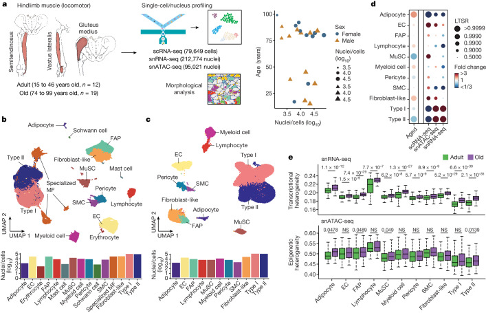
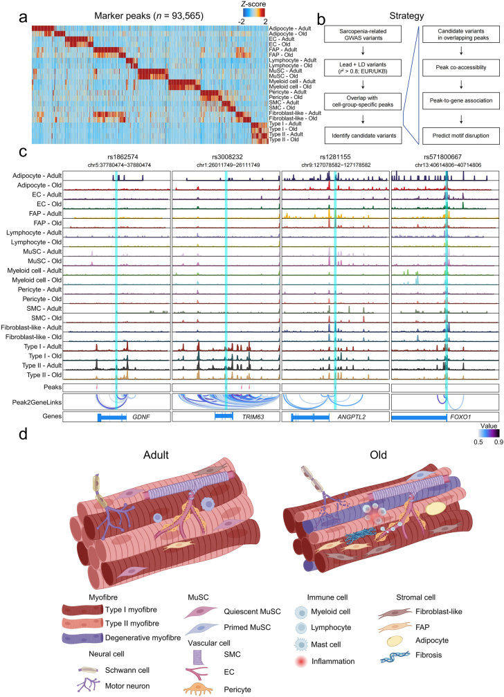
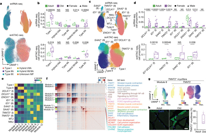
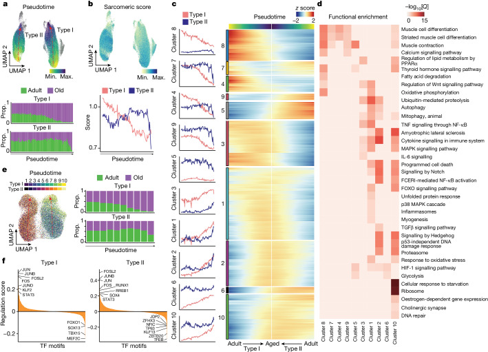
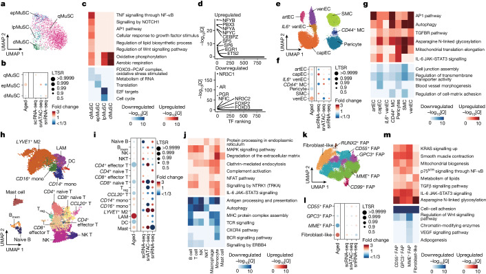
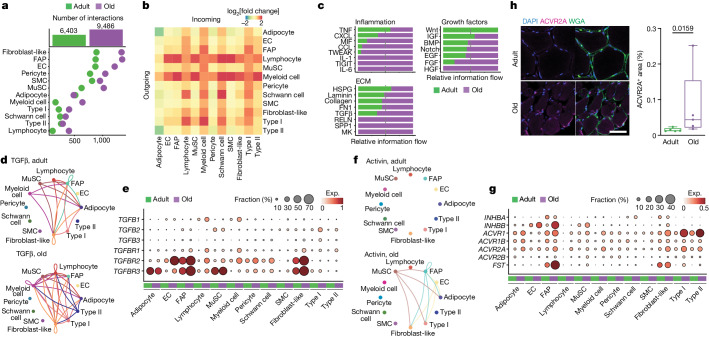
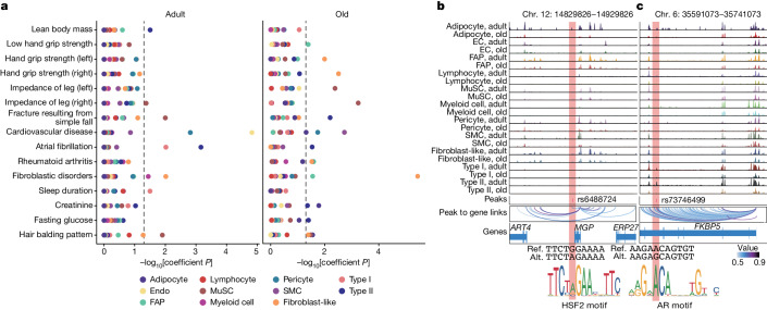
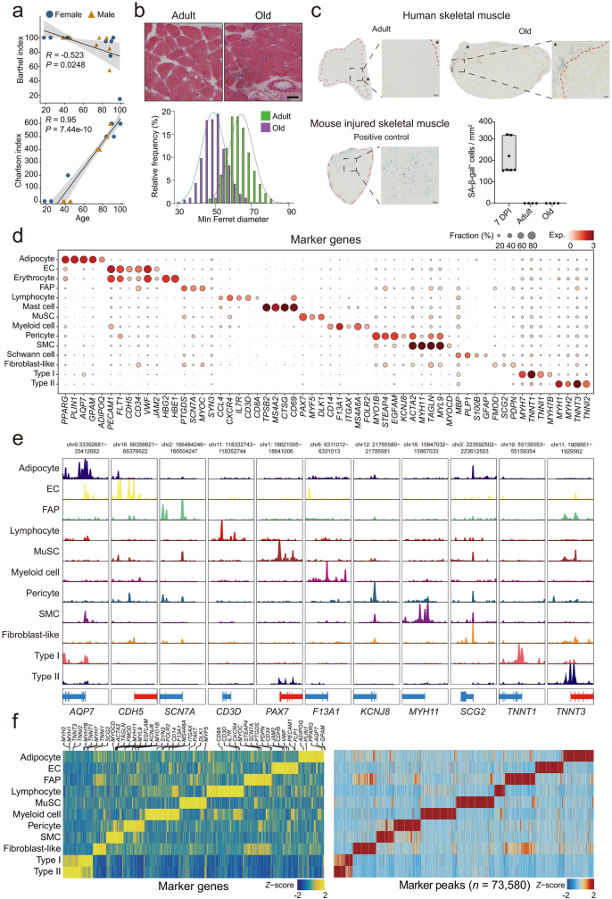
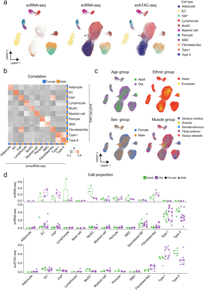
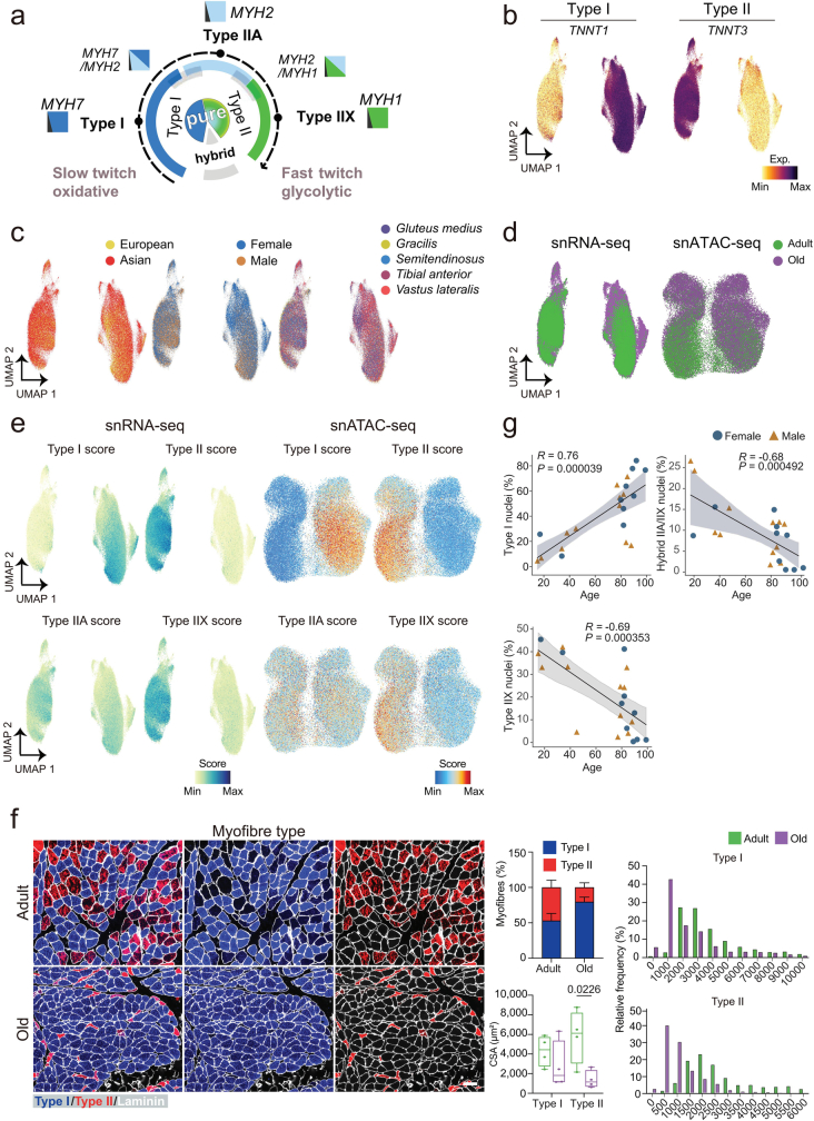
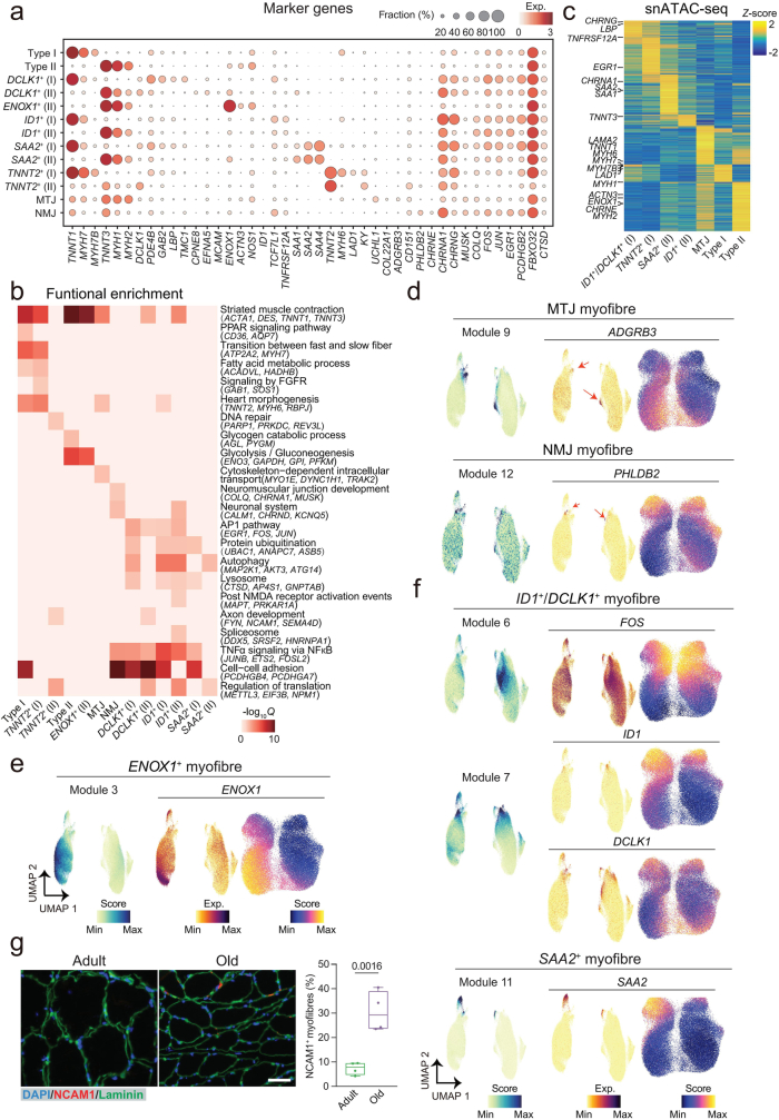
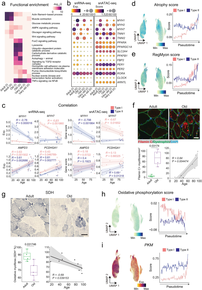
Muscle atrophy and functional decline (sarcopenia) are common manifestations of frailty and are critical contributors to morbidity and mortality in older people. Deciphering the molecular mechanisms underlying sarcopenia has major implications for understanding human ageing. Yet, progress has been slow, partly due to the difficulties of characterizing skeletal muscle niche heterogeneity (whereby myofibres are the most abundant) and obtaining well-characterized human samples. Here we generate a single-cell/single-nucleus transcriptomic and chromatin accessibility map of human limb skeletal muscles encompassing over 387,000 cells/nuclei from individuals aged 15 to 99 years with distinct fitness and frailty levels. We describe how cell populations change during ageing, including the emergence of new populations in older people, and the cell-specific and multicellular network features (at the transcriptomic and epigenetic levels) associated with these changes. On the basis of cross-comparison with genetic data, we also identify key elements of chromatin architecture that mark susceptibility to sarcopenia. Our study provides a basis for identifying targets in the skeletal muscle that are amenable to medical, pharmacological and lifestyle interventions in late life.

摘要

肌肉萎缩和功能下降(肌少症)是衰弱的常见表现,也是老年人发病率和死亡率的重要原因。解析肌少症的分子机制对于理解人类衰老具有重要意义。然而,进展缓慢,部分原因是难以描述骨骼肌肉生态位异质性(其中肌纤维最为丰富)和获得特征明确的人类样本。在这里,我们生成了一个单细胞/单核转录组和染色质可及性图谱,涵盖了来自年龄在 15 岁至 99 岁之间、具有不同健康水平和衰弱程度的个体的超过 387000 个细胞/核。我们描述了细胞群体在衰老过程中的变化,包括在老年人中出现的新群体,以及与这些变化相关的细胞特异性和多细胞网络特征(在转录组和表观遗传水平上)。基于与遗传数据的交叉比较,我们还确定了染色质结构的关键元素,这些元素标志着对肌少症的易感性。我们的研究为确定骨骼肌肉中的靶标提供了基础,这些靶标可以通过医学、药理学和生活方式干预来治疗晚年的肌少症。

https://cdn.ncbi.nlm.nih.gov/pmc/blobs/50c1/11062927/55c20a45f9d8/41586_2024_7348_Fig19_ESM.jpg
https://cdn.ncbi.nlm.nih.gov/pmc/blobs/50c1/11062927/76526f9e4e33/41586_2024_7348_Fig1_HTML.jpg
https://cdn.ncbi.nlm.nih.gov/pmc/blobs/50c1/11062927/4a3016bd887f/41586_2024_7348_Fig2_HTML.jpg
https://cdn.ncbi.nlm.nih.gov/pmc/blobs/50c1/11062927/f4dbe7a1621d/41586_2024_7348_Fig3_HTML.jpg
https://cdn.ncbi.nlm.nih.gov/pmc/blobs/50c1/11062927/f3b6071c7c64/41586_2024_7348_Fig4_HTML.jpg
https://cdn.ncbi.nlm.nih.gov/pmc/blobs/50c1/11062927/e7ba633f6748/41586_2024_7348_Fig5_HTML.jpg
https://cdn.ncbi.nlm.nih.gov/pmc/blobs/50c1/11062927/282d7f0fb5c1/41586_2024_7348_Fig6_HTML.jpg
https://cdn.ncbi.nlm.nih.gov/pmc/blobs/50c1/11062927/f8fd5b3c20c1/41586_2024_7348_Fig7_ESM.jpg
https://cdn.ncbi.nlm.nih.gov/pmc/blobs/50c1/11062927/67a3cef200e2/41586_2024_7348_Fig8_ESM.jpg
https://cdn.ncbi.nlm.nih.gov/pmc/blobs/50c1/11062927/fe89b0d42d86/41586_2024_7348_Fig9_ESM.jpg
https://cdn.ncbi.nlm.nih.gov/pmc/blobs/50c1/11062927/108ad693c897/41586_2024_7348_Fig10_ESM.jpg
https://cdn.ncbi.nlm.nih.gov/pmc/blobs/50c1/11062927/10ada9b2c526/41586_2024_7348_Fig11_ESM.jpg
https://cdn.ncbi.nlm.nih.gov/pmc/blobs/50c1/11062927/396308c1012d/41586_2024_7348_Fig12_ESM.jpg
https://cdn.ncbi.nlm.nih.gov/pmc/blobs/50c1/11062927/edfb6f0c934f/41586_2024_7348_Fig13_ESM.jpg
https://cdn.ncbi.nlm.nih.gov/pmc/blobs/50c1/11062927/40bd6a2bad68/41586_2024_7348_Fig14_ESM.jpg
https://cdn.ncbi.nlm.nih.gov/pmc/blobs/50c1/11062927/6488fb840e6b/41586_2024_7348_Fig15_ESM.jpg
https://cdn.ncbi.nlm.nih.gov/pmc/blobs/50c1/11062927/4514fa7ab2aa/41586_2024_7348_Fig16_ESM.jpg
https://cdn.ncbi.nlm.nih.gov/pmc/blobs/50c1/11062927/d43038ff01b9/41586_2024_7348_Fig17_ESM.jpg
https://cdn.ncbi.nlm.nih.gov/pmc/blobs/50c1/11062927/7dbf89ed3c8f/41586_2024_7348_Fig18_ESM.jpg
https://cdn.ncbi.nlm.nih.gov/pmc/blobs/50c1/11062927/55c20a45f9d8/41586_2024_7348_Fig19_ESM.jpg

相似文献

1
Multimodal cell atlas of the ageing human skeletal muscle.衰老人体骨骼肌的多模态细胞图谱。
Nature. 2024 May;629(8010):154-164. doi: 10.1038/s41586-024-07348-6. Epub 2024 Apr 22.
2
Basic Science of Frailty-Biological Mechanisms of Age-Related Sarcopenia.虚弱的基础科学-与年龄相关的肌肉减少症的生物学机制。
Anesth Analg. 2021 Feb 1;132(2):293-304. doi: 10.1213/ANE.0000000000005096.
3
A robust neuromuscular system protects rat and human skeletal muscle from sarcopenia.一个强健的神经肌肉系统可保护大鼠和人类骨骼肌免受肌肉减少症的影响。
Aging (Albany NY). 2016 Apr;8(4):712-29. doi: 10.18632/aging.100926.
4
Characterizing the skeletal muscle immune microenvironment for sarcopenia: insights from transcriptome analysis and histological validation.从转录组分析和组织学验证角度探讨骨骼肌免疫微环境与肌肉减少症的关系
Front Immunol. 2024 Jul 4;15:1414387. doi: 10.3389/fimmu.2024.1414387. eCollection 2024.
5
Aging- and obesity-related peri-muscular adipose tissue accelerates muscle atrophy.衰老和肥胖相关的肌周脂肪组织加速肌肉萎缩。
PLoS One. 2019 Aug 23;14(8):e0221366. doi: 10.1371/journal.pone.0221366. eCollection 2019.
6
Nrf2 deficiency exacerbates frailty and sarcopenia by impairing skeletal muscle mitochondrial biogenesis and dynamics in an age-dependent manner.Nrf2 缺乏通过损害骨骼肌线粒体生物发生和动态平衡以年龄依赖的方式加剧虚弱和肌肉减少症。
Exp Gerontol. 2019 May;119:61-73. doi: 10.1016/j.exger.2019.01.022. Epub 2019 Jan 25.
7
Overweight in elderly people induces impaired autophagy in skeletal muscle.老年人超重会导致骨骼肌自噬受损。
Free Radic Biol Med. 2017 Sep;110:31-41. doi: 10.1016/j.freeradbiomed.2017.05.018. Epub 2017 May 23.
8
Ageing in relation to skeletal muscle dysfunction: redox homoeostasis to regulation of gene expression.与骨骼肌功能障碍相关的衰老:从氧化还原稳态到基因表达调控
Mamm Genome. 2016 Aug;27(7-8):341-57. doi: 10.1007/s00335-016-9643-x. Epub 2016 May 23.
9
Integrated genomic and proteomic analyses identify stimulus-dependent molecular changes associated with distinct modes of skeletal muscle atrophy.整合基因组和蛋白质组分析鉴定了与不同类型骨骼肌萎缩相关的刺激依赖性分子变化。
Cell Rep. 2021 Nov 9;37(6):109971. doi: 10.1016/j.celrep.2021.109971.
10
Apoptosis in capillary endothelial cells in ageing skeletal muscle.衰老骨骼肌中毛细血管内皮细胞的凋亡
Aging Cell. 2014 Apr;13(2):254-62. doi: 10.1111/acel.12169. Epub 2013 Nov 19.

引用本文的文献

1
Epigenetic Regulation of Aging and its Rejuvenation.衰老及其逆转的表观遗传调控
MedComm (2020). 2025 Sep 1;6(9):e70369. doi: 10.1002/mco2.70369. eCollection 2025 Sep.
2
Sarcopenia index based on serum creatinine and cystatin C is associated with the risk of stroke in middle-aged and older adults in Chinese: a prospective cohort study from the China health and retirement longitudinal study.基于血清肌酐和胱抑素C的肌少症指数与中国中老年人群的中风风险相关:一项来自中国健康与养老追踪调查的前瞻性队列研究
BMC Neurol. 2025 Sep 1;25(1):371. doi: 10.1186/s12883-025-04327-w.
3
Multi-omic integration of single-cell data uncovers methylation profiles of super-enhancers in skeletal muscle stem cells.

本文引用的文献

1
Sarcopenia: no consensus, no diagnostic criteria, and no approved indication-How did we get here?肌肉减少症:尚无共识,无诊断标准,也无获批适应证——我们是如何走到这一步的?
Geroscience. 2024 Feb;46(1):183-190. doi: 10.1007/s11357-023-01016-9. Epub 2023 Nov 24.
2
Transcriptional activation of Jun and Fos members of the AP-1 complex is a conserved signature of immune aging that contributes to inflammaging.AP-1 复合物中 Jun 和 Fos 成员的转录激活是免疫衰老的保守特征,有助于炎症衰老。
Aging Cell. 2023 Apr;22(4):e13792. doi: 10.1111/acel.13792. Epub 2023 Feb 24.
3
MME fibro-adipogenic progenitors are the dominant adipogenic population during fatty infiltration in human skeletal muscle.
单细胞数据的多组学整合揭示了骨骼肌干细胞中超增强子的甲基化谱。
Epigenetics Chromatin. 2025 Aug 11;18(1):54. doi: 10.1186/s13072-025-00619-0.
4
Large MAF transcription factors reawaken evolutionarily dormant fast-glycolytic type IIb myofibers in human skeletal muscle.大型MAF转录因子可使人类骨骼肌中进化上处于休眠状态的快速糖酵解IIb型肌纤维重新激活。
Skelet Muscle. 2025 Jul 26;15(1):19. doi: 10.1186/s13395-025-00391-5.
5
Mechanical effects in aging of the musculoskeletal system: Molecular signaling and spatial scale alterations.肌肉骨骼系统衰老中的力学效应:分子信号传导与空间尺度改变
J Orthop Translat. 2025 May 27;52:464-477. doi: 10.1016/j.jot.2025.04.018. eCollection 2025 May.
6
Addressing osteoblast senescence: Molecular pathways and the frontier of anti-ageing treatments.应对成骨细胞衰老:分子途径与抗衰老治疗前沿
Clin Transl Med. 2025 Jul;15(7):e70417. doi: 10.1002/ctm2.70417.
7
Molecular constraints of sarcopenia in the ageing muscle.衰老肌肉中肌肉减少症的分子限制因素
Front Aging. 2025 Jul 3;6:1588014. doi: 10.3389/fragi.2025.1588014. eCollection 2025.
8
Transcriptional Diversity in Response to Aging Across Skeletal Muscles.衰老过程中不同骨骼肌的转录多样性
Aging Cell. 2025 Sep;24(9):e70164. doi: 10.1111/acel.70164. Epub 2025 Jul 9.
9
Genomics reveals eleven obesity endotypes with distinct biological and phenotypic signatures.基因组学揭示了具有不同生物学和表型特征的11种肥胖内型。
medRxiv. 2025 Jul 2:2025.06.30.25330607. doi: 10.1101/2025.06.30.25330607.
10
Multiomics and cellular senescence profiling of aging human skeletal muscle uncovers Maraviroc as a senotherapeutic approach for sarcopenia.衰老人类骨骼肌的多组学和细胞衰老分析揭示马拉维若可作为一种治疗肌肉减少症的衰老疗法。
Nat Commun. 2025 Jul 5;16(1):6207. doi: 10.1038/s41467-025-61403-y.
MME 纤维脂肪祖细胞是人骨骼肌脂肪浸润过程中主要的成脂细胞群体。
Commun Biol. 2023 Jan 27;6(1):111. doi: 10.1038/s42003-023-04504-y.
4
Defining the age-dependent and tissue-specific circadian transcriptome in male mice.定义雄性小鼠中与年龄相关和组织特异性的昼夜转录组。
Cell Rep. 2023 Jan 31;42(1):111982. doi: 10.1016/j.celrep.2022.111982. Epub 2023 Jan 9.
5
Senescence atlas reveals an aged-like inflamed niche that blunts muscle regeneration.衰老图谱揭示了一种衰老样炎症微环境,削弱了肌肉再生。
Nature. 2023 Jan;613(7942):169-178. doi: 10.1038/s41586-022-05535-x. Epub 2022 Dec 21.
6
Single nuclei profiling identifies cell specific markers of skeletal muscle aging, frailty, and senescence.单细胞分析鉴定骨骼肌衰老、虚弱和衰老的细胞特异性标志物。
Aging (Albany NY). 2022 Dec 13;14(23):9393-9422. doi: 10.18632/aging.204435.
7
Inflammaging: Implications in Sarcopenia.慢性炎症与肌肉减少症
Int J Mol Sci. 2022 Nov 30;23(23):15039. doi: 10.3390/ijms232315039.
8
Functional inference of gene regulation using single-cell multi-omics.利用单细胞多组学进行基因调控的功能推断
Cell Genom. 2022 Sep 14;2(9). doi: 10.1016/j.xgen.2022.100166. Epub 2022 Aug 4.
9
The FASTQ+ format and PISA.FASTQ+ 格式和 PISA。
Bioinformatics. 2022 Sep 30;38(19):4639-4642. doi: 10.1093/bioinformatics/btac562.
10
Inhibition of TNF-α Restores Muscle Force, Inhibits Inflammation, and Reduces Apoptosis of Traumatized Skeletal Muscles.抑制 TNF-α 可恢复肌肉力量,抑制炎症,并减少创伤性骨骼肌的细胞凋亡。
Cells. 2022 Aug 3;11(15):2397. doi: 10.3390/cells11152397.