VIB Center for Inflammation Research, 9052, Ghent, Belgium.
Department of Biomedical Molecular Biology, Ghent University, 9052, Ghent, Belgium.
Cell Death Differ. 2024 Aug;31(8):957-969. doi: 10.1038/s41418-024-01297-3. Epub 2024 Apr 22.
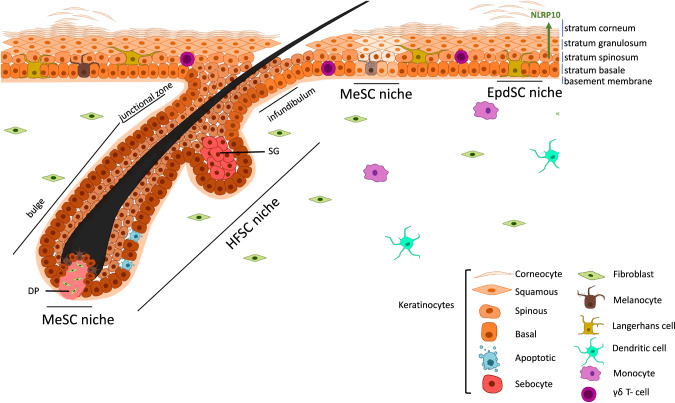
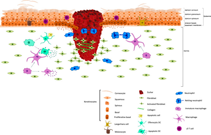
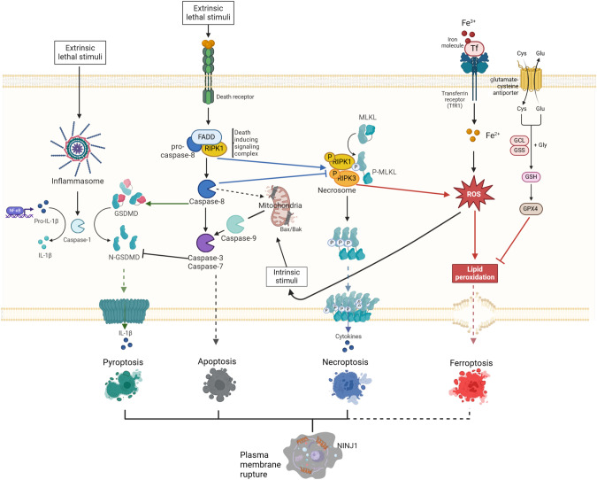
Our skin provides a physical and immunological barrier against dehydration and environmental insults ranging from microbial attacks, toxins and UV irradiation to wounding. Proper functioning of the skin barrier largely depends on the interplay between keratinocytes- the epithelial cells of the skin- and immune cells. Two spatially distinct populations of keratinocyte stem cells (SCs) maintain the epidermal barrier function and the hair follicle. These SCs are inherently long-lived, but cell death can occur within their niches and impacts their functionality. The default cell death programme in skin is apoptosis, an orderly and non-inflammatory suicide programme. However, recent findings are shedding light on the significance of various modes of regulated necrotic cell death, which are lytic and can provoke inflammation within the local skin environment. While the presence of dying cells was generally regarded as a mere consequence of inflammation, findings in various human dermatological conditions and experimental mouse models of aberrant cell death control demonstrated that cell death programmes in keratinocytes (KCs) can drive skin inflammation and even tumour initiation. When cells die, they need to be removed by phagocytosis and KCs can function as non-professional phagocytes of apoptotic cells with important implications for their SC capacities. It is becoming apparent that in conditions of heightened SC activity, distinct cell death modalities differentially impact the different skin SC populations in their local niches. Here, we describe how regulated cell death modalities functionally affect epidermal SC niches along with their relevance to injury repair, inflammatory skin disorders and cancer.
我们的皮肤提供了物理和免疫屏障,防止脱水和环境侵害,包括微生物攻击、毒素和紫外线辐射以及创伤。皮肤屏障的正常功能在很大程度上取决于角质形成细胞(皮肤的上皮细胞)和免疫细胞之间的相互作用。两种空间上不同的角质形成细胞干细胞(SCs)群体维持着表皮屏障功能和毛囊。这些SCs 具有固有长寿命,但细胞死亡可能发生在其龛位内,并影响其功能。皮肤中默认的细胞死亡程序是细胞凋亡,这是一种有序的、非炎症性的自杀程序。然而,最近的发现揭示了各种受调控的坏死性细胞死亡方式的重要性,这些方式是溶酶体的,可以在局部皮肤环境中引发炎症。虽然死亡细胞的存在通常被认为仅仅是炎症的结果,但在各种人类皮肤病和异常细胞死亡控制的实验小鼠模型中发现,角质形成细胞(KCs)中的细胞死亡程序可以驱动皮肤炎症,甚至启动肿瘤。当细胞死亡时,它们需要通过吞噬作用被清除,而 KCs 可以作为凋亡细胞的非专业吞噬细胞发挥作用,这对其 SC 能力有重要影响。越来越明显的是,在 SC 活性增强的情况下,不同的细胞死亡方式会以不同的方式影响局部龛位中的不同皮肤 SC 群体。在这里,我们描述了受调控的细胞死亡方式如何影响表皮 SC 龛位,以及它们与损伤修复、炎症性皮肤疾病和癌症的相关性。