Jiang Chen, Ma Cai Y, Hazlehurst Thomas A, Ilett Thomas P, Jackson Alexander S M, Hogg David C, Roberts Kevin J
Centre for the Digital Design of Drug Products, School of Chemical and Process Engineering, University of Leeds, Leeds LS2 9JT, U.K.
School of Computing, University of Leeds, Leeds LS2 9JT, U.K.
Cryst Growth Des. 2024 Apr 5;24(8):3277-3288. doi: 10.1021/acs.cgd.3c01548. eCollection 2024 Apr 17.
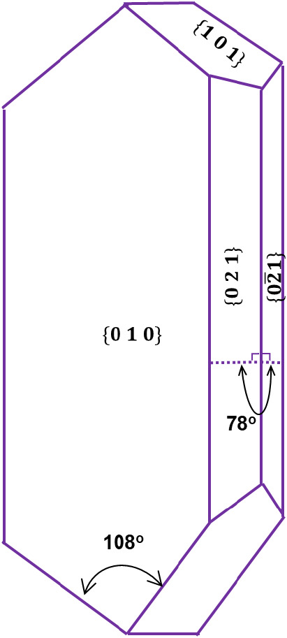
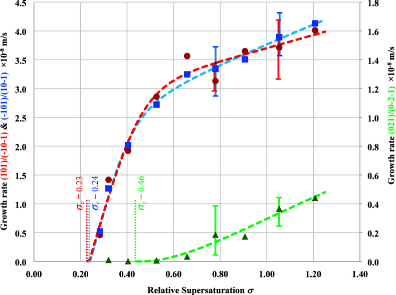
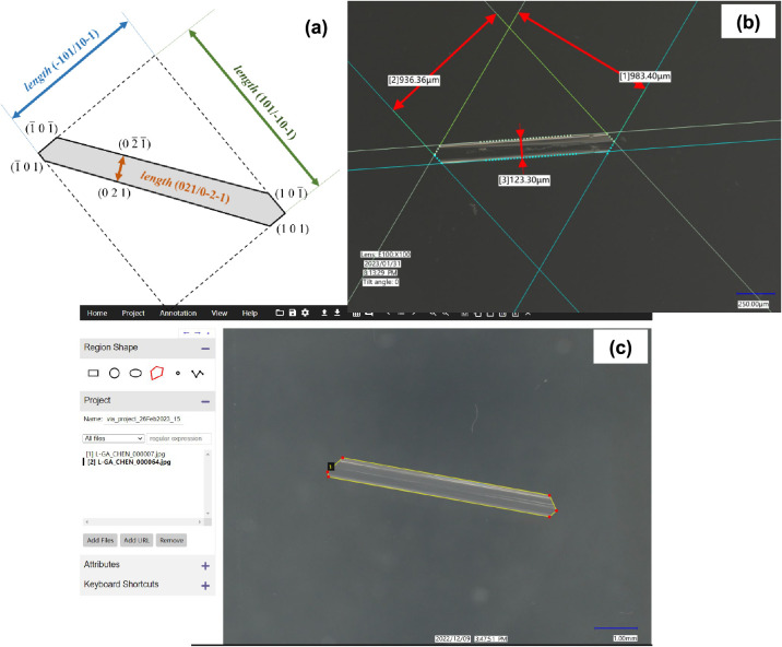
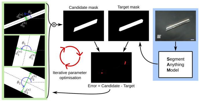
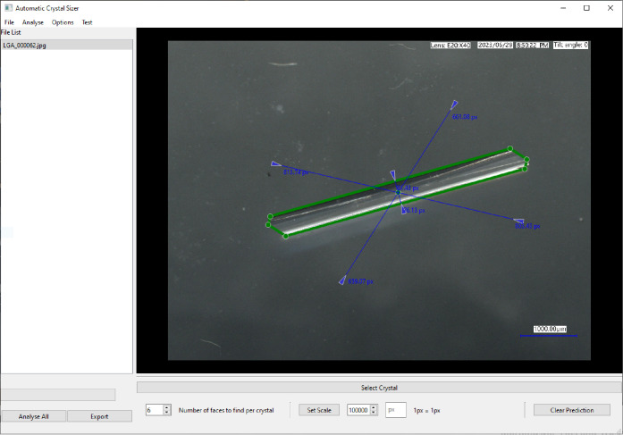
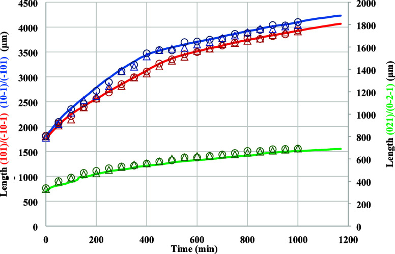
Precision measurement of the growth rate of individual single crystal facets () represents an important component in the design of industrial crystallization processes. Current approaches for crystal growth measurement using optical microscopy are labor intensive and prone to error. An automated process using state-of-the-art computer vision and machine learning to segment and measure the crystal images is presented. The accuracies and efficiencies of the new crystal sizing approach are evaluated against existing manual and semi-automatic methods, demonstrating equivalent accuracy but over a much shorter time, thereby enabling a more complete kinematic analysis of the overall crystallization process. This is applied to measure in situ the crystal growth rates and through this determining the associated kinetic mechanisms for the crystallization of β-form l-glutamic acid from the solution phase. Growth on the {101} capping faces is consistent with a Birth and Spread mechanism, in agreement with the literature, while the growth rate of the {021} prismatic faces, previously not available in the literature, is consistent with a Burton-Cabrera-Frank screw dislocation mechanism. At a typical supersaturation of σ = 0.78, the growth rate of the {101} capping faces (3.2 × 10 m s) is found to be 17 times that of the {021} prismatic faces (1.9 × 10 m s). Both capping and prismatic faces are found to have dead zones in their growth kinetic profiles, with the capping faces ( = 0.23) being about half that of the prismatic faces ( = 0.46). The importance of this overall approach as an integral component of the digital design of industrial crystallization processes is highlighted.
精确测量单个单晶面的生长速率()是工业结晶过程设计中的一个重要组成部分。目前使用光学显微镜进行晶体生长测量的方法劳动强度大且容易出错。本文提出了一种利用先进的计算机视觉和机器学习对晶体图像进行分割和测量的自动化方法。将这种新的晶体尺寸测量方法的精度和效率与现有的手动和半自动方法进行了评估,结果表明其精度相当,但所需时间更短,从而能够对整个结晶过程进行更完整的运动学分析。这一方法被应用于原位测量晶体生长速率,并由此确定从溶液相中结晶出β型L-谷氨酸的相关动力学机制。{101}封端面的生长符合成核与扩展机制,与文献一致,而{021}棱柱面的生长速率(此前文献中未报道)符合伯顿-卡布雷拉-弗兰克螺旋位错机制。在典型过饱和度σ = 0.78时,发现{101}封端面的生长速率(3.2×10 m s)是{021}棱柱面(1.9×10 m s)的17倍。发现封端面和棱柱面在其生长动力学曲线上都有死区,封端面的死区( = 0.23)约为棱柱面( = 0.46)的一半。强调了这种整体方法作为工业结晶过程数字设计不可或缺的组成部分的重要性。