Department of Biomedical Imaging and Image-Guided Therapy, Division of Nuclear Medicine, Medical University of Vienna, Währinger Gürtel 18-20, 1090, Vienna, Austria.
Christian Doppler Laboratory for Applied Metabolomics (CDL AM), Medical University of Vienna, Vienna, Austria.
Eur J Nucl Med Mol Imaging. 2024 Jul;51(9):2833-2842. doi: 10.1007/s00259-024-06698-7. Epub 2024 May 2.
Circulating-tumor DNA (ctDNA) and prostate-specific membrane antigen (PSMA) ligand positron-emission tomography (PET) enable minimal-invasive prostate cancer (PCa) detection and survival prognostication. The present study aims to compare their tumor discovery abilities and prognostic values.
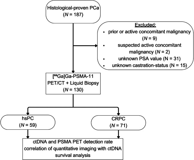
One hundred thirty men with confirmed PCa (70.5 ± 8.0 years) who underwent [Ga]Ga-PSMA-11 PET/CT (184.8 ± 19.7 MBq) imaging and plasma sample collection (March 2019-August 2021) were included. Plasma-extracted cell-free DNA was subjected to whole-genome-based ctDNA analysis. PSMA-positive tumor lesions were delineated and their quantitative parameters extracted. ctDNA and PSMA PET/CT discovery rates were compared, and the prognostic value for overall survival (OS) was evaluated.
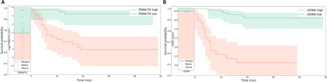
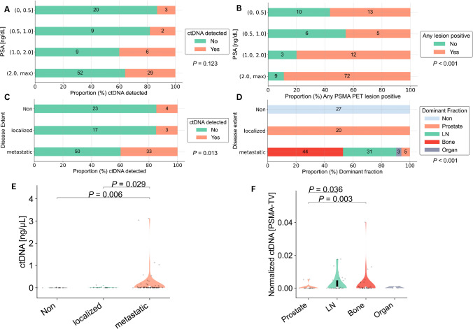
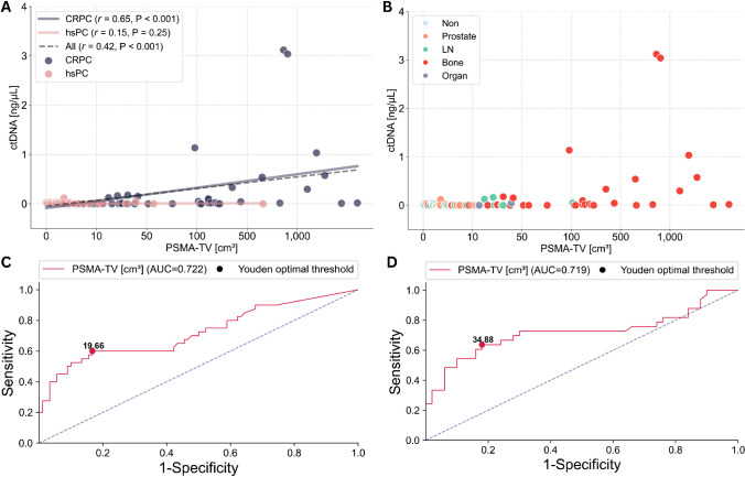
PSMA PET discovery rates according to castration status and PSA ranges did differ significantly (P = 0.013, P < 0.001), while ctDNA discovery rates did not (P = 0.311, P = 0.123). ctDNA discovery rates differed between localized and metastatic disease (P = 0.013). Correlations between ctDNA concentrations and PSMA-positive tumor volume (PSMA-TV) were significant in all (r = 0.42, P < 0.001) and castration-resistant (r = 0.65, P < 0.001), however not in hormone-sensitive patients (r = 0.15, P = 0.249). PSMA-TV and ctDNA levels were associated with survival outcomes in the Logrank (P < 0.0001, P < 0.0001) and multivariate Cox regression analysis (P = 0.0023, P < 0.0001).
These findings suggest that PSMA PET imaging outperforms ctDNA analysis in detecting prostate cancer across the whole spectrum of disease, while both modalities are independently highly prognostic for survival outcomes.
循环肿瘤 DNA(ctDNA)和前列腺特异性膜抗原(PSMA)配体正电子发射断层扫描(PET)能够实现对前列腺癌(PCa)的微创检测和生存预后预测。本研究旨在比较它们的肿瘤发现能力和预后价值。
本研究共纳入 130 名经确诊患有 PCa 的男性患者(70.5±8.0 岁),他们于 2019 年 3 月至 2021 年 8 月期间接受了[Ga]Ga-PSMA-11 PET/CT(184.8±19.7MBq)成像和血浆样本采集。从血浆中提取的无细胞 DNA 进行全基因组 ctDNA 分析。勾画 PSMA 阳性肿瘤病灶并提取其定量参数。比较 ctDNA 和 PSMA PET/CT 的检出率,并评估其对总生存(OS)的预后价值。
根据去势状态和 PSA 范围,PSMA PET 的检出率存在显著差异(P=0.013,P<0.001),而 ctDNA 的检出率无差异(P=0.311,P=0.123)。ctDNA 的检出率在局限性疾病和转移性疾病之间存在差异(P=0.013)。ctDNA 浓度与 PSMA 阳性肿瘤体积(PSMA-TV)之间存在显著相关性,在所有患者(r=0.42,P<0.001)和去势抵抗患者(r=0.65,P<0.001)中均如此,但在激素敏感患者中则不然(r=0.15,P=0.249)。PSMA-TV 和 ctDNA 水平在 Logrank 检验(P<0.0001,P<0.0001)和多变量 Cox 回归分析(P=0.0023,P<0.0001)中与生存结果相关。
这些发现表明,PSMA PET 成像在检测整个疾病谱中的前列腺癌方面优于 ctDNA 分析,而这两种方法在预测生存结果方面均具有高度的独立性。