Department of Radiology and Biomedical Imaging, University of California San Francisco, San Francisco, California.
Helen Diller Comprehensive Cancer Center, University of California San Francisco, San Francisco, California.
J Nucl Med. 2024 Jun 3;65(6):938-943. doi: 10.2967/jnumed.123.267281.
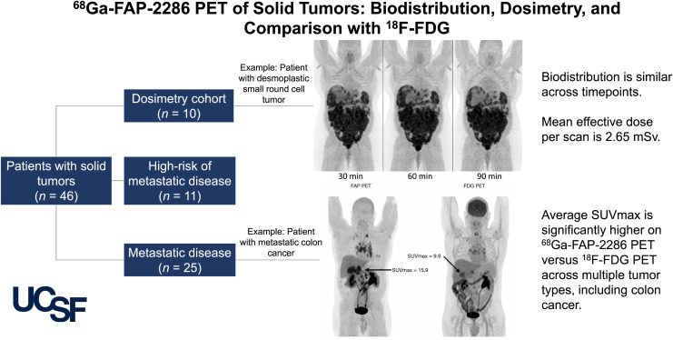
Fibroblast activation protein (FAP), expressed in the tumor microenvironment of a variety of cancers, has become a target of novel PET tracers. The purpose of this report is to evaluate the imaging characteristics of Ga-FAP-2286, present the first-to our knowledge-dosimetry analysis to date, and compare the agent with F-FDG and FAPI compounds. Patients were administered 219 ± 43 MBq of Ga-FAP-2286 and scanned after 60 min. Uptake was measured in up to 5 lesions per patient and within the kidneys, spleen, liver, and mediastinum (blood pool). Absorbed doses were evaluated using MIM Encore and OLINDA/EXM version 1.1 using the International Commission on Radiological Protection publication 103 tissue weighting factor. Forty-six patients were imaged with Ga-FAP-2286 PET. The highest average uptake was seen in sarcoma, cholangiocarcinoma, and colon cancer. The lowest uptake was found in lung cancer and testicular cancer. The average SUV was significantly higher on Ga-FAP-2286 PET than on F-FDG PET in cholangiocarcinoma (18.2 ± 6.4 vs. 9.1 ± 5.0, = 0.007), breast cancer (11.1 ± 6.8 vs. 4.1 ± 2.2, < 0.001), colon cancer (13.8 ± 2.2 vs. 7.6 ± 1.7, = 0.001), hepatocellular carcinoma (9.3 ± 3.5 vs. 4.7 ± 1.3, = 0.01), head and neck cancer (11.3 ± 3.5 vs. 7.6 ± 5.5, = 0.04), and pancreatic adenocarcinoma (7.4 ± 1.8 vs. 3.7 ± 1.0, = 0.01). The total-body effective dose was estimated at 1.16E-02 mSv/MBq, with the greatest absorbed organ dose in the urinary bladder wall (9.98E-02 mGy/MBq). Ga-FAP-2286 biodistribution, dosimetry, and tumor uptake were similar to those of previously reported FAPI compounds. Additionally,Ga-FAP-2286 PET had consistently higher uptake than F-FDG PET. These results are especially promising in the setting of small-volume disease and differentiating tumor from inflammatory uptake.
成纤维细胞激活蛋白(FAP)在多种癌症的肿瘤微环境中表达,已成为新型 PET 示踪剂的靶标。本报告的目的是评估 Ga-FAP-2286 的成像特征,报告迄今为止首次的药物代谢动力学分析,并比较该示踪剂与 F-FDG 和 FAPI 化合物。每位患者给予 219±43MBq 的 Ga-FAP-2286,并在 60 分钟后进行扫描。在每位患者最多 5 个病灶内以及肾脏、脾脏、肝脏和纵隔(血池)内测量摄取量。使用 MIM Encore 和 OLINDA/EXM 版本 1.1 以及国际辐射防护委员会出版物 103 组织权重因子评估吸收剂量。46 名患者接受了 Ga-FAP-2286 PET 成像。肉瘤、胆管癌和结肠癌的摄取量最高。肺癌和睾丸癌的摄取量最低。胆管癌(18.2±6.4 比 9.1±5.0, = 0.007)、乳腺癌(11.1±6.8 比 4.1±2.2, < 0.001)、结肠癌(13.8±2.2 比 7.6±1.7, = 0.001)、肝细胞癌(9.3±3.5 比 4.7±1.3, = 0.01)、头颈部癌(11.3±3.5 比 7.6±5.5, = 0.04)和胰腺腺癌(7.4±1.8 比 3.7±1.0, = 0.01)。Ga-FAP-2286 的全身有效剂量估计为 1.16E-02mSv/MBq,膀胱壁的吸收器官剂量最大(9.98E-02mGy/MBq)。Ga-FAP-2286 的生物分布、药物代谢动力学和肿瘤摄取与之前报道的 FAPI 化合物相似。此外,Ga-FAP-2286 PET 的摄取量始终高于 F-FDG PET。这些结果在小体积疾病和区分肿瘤与炎症摄取方面尤其有希望。