Maity Sudipa, Huang Yuanyu, Kilgore Mitchell D, Thurmon Abbigail N, Vaasjo Lee O, Galazo Maria J, Xu Xiaojiang, Cao Jing, Wang Xiaoying, Ning Bo, Liu Ning, Fan Jia
Center for Cellular and Molecular Diagnostics, Tulane University School of Medicine, New Orleans, LA, USA.
Department of Biochemistry and Molecular Biology, Tulane University School of Medicine, New Orleans, LA, USA.
Clin Proteomics. 2024 May 12;21(1):32. doi: 10.1186/s12014-024-09485-6.
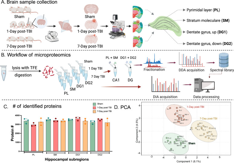
Traumatic brain injury (TBI) often results in diverse molecular responses, challenging traditional proteomic studies that measure average changes at tissue levels and fail to capture the complexity and heterogeneity of the affected tissues. Spatial proteomics offers a solution by providing insights into sub-region-specific alterations within tissues. This study focuses on the hippocampal sub-regions, analyzing proteomic expression profiles in mice at the acute (1 day) and subacute (7 days) phases of post-TBI to understand subregion-specific vulnerabilities and long-term consequences.
Three mice brains were collected from each group, including Sham, 1-day post-TBI and 7-day post-TBI. Hippocampal subregions were extracted using Laser Microdissection (LMD) and subsequently analyzed by label-free quantitative proteomics.
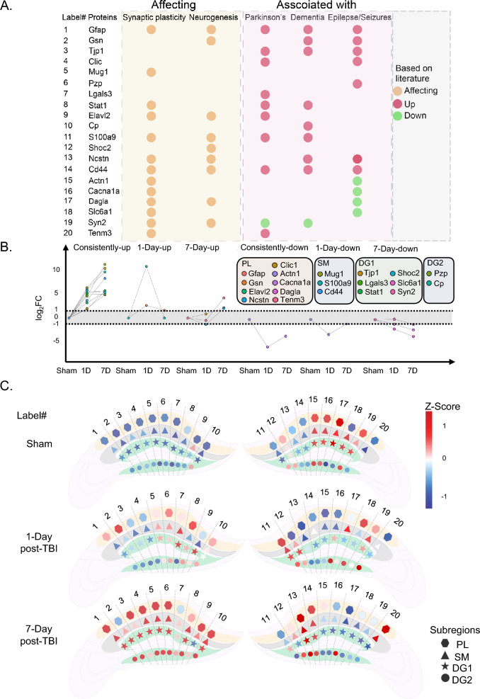
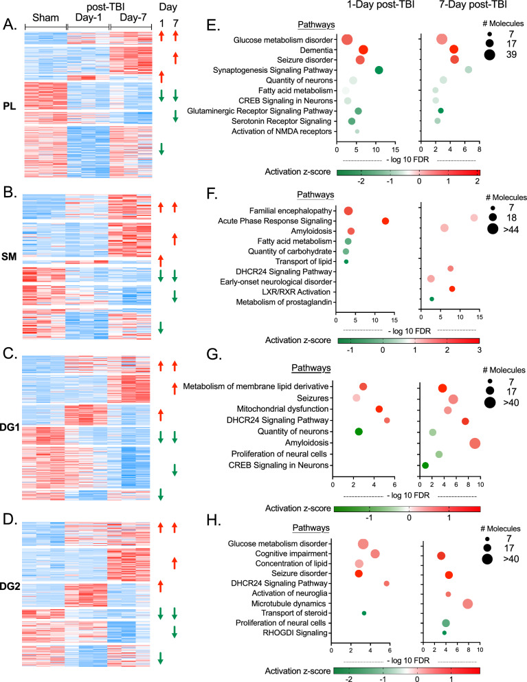
The spatial analysis reveals region-specific protein abundance changes, highlighting the elevation of FN1, LGALS3BP, HP, and MUG-1 in the stratum moleculare (SM), suggesting potential immune cell enrichment post-TBI. Notably, established markers of chronic traumatic encephalopathy, IGHM and B2M, exhibit specific upregulation in the dentate gyrus bottom (DG2) independent of direct mechanical injury. Metabolic pathway analysis identifies disturbances in glucose and lipid metabolism, coupled with activated cholesterol synthesis pathways enriched in SM at 7-Day post-TBI and subsequently in deeper DG1 and DG2 suggesting a role in neurogenesis and the onset of recovery. Coordinated activation of neuroglia and microtubule dynamics in DG2 suggest recovery mechanisms in less affected regions. Cluster analysis revealed spatial variations post-TBI, indicative of dysregulated neuronal plasticity and neurogenesis and further predisposition to neurological disorders. TBI-induced protein upregulation (MUG-1, PZP, GFAP, TJP, STAT-1, and CD44) across hippocampal sub-regions indicates shared molecular responses and links to neurological disorders. Spatial variations were demonstrated by proteins dysregulated in both or either of the time-points exclusively in each subregion (ELAVL2, CLIC1 in PL, CD44 and MUG-1 in SM, and SHOC2, LGALS3 in DG).
Utilizing advanced spatial proteomics techniques, the study unveils the dynamic molecular responses in distinct hippocampal subregions post-TBI. It uncovers region-specific vulnerabilities and dysregulated neuronal processes, and potential recovery-related pathways that contribute to our understanding of TBI's neurological consequences and provides valuable insights for biomarker discovery and therapeutic targets.
创伤性脑损伤(TBI)常引发多种分子反应,这给传统蛋白质组学研究带来挑战,传统研究只能测量组织水平的平均变化,无法捕捉受影响组织的复杂性和异质性。空间蛋白质组学通过深入了解组织内特定亚区域的变化提供了解决方案。本研究聚焦于海马亚区域,分析TBI后急性(1天)和亚急性(7天)阶段小鼠的蛋白质组表达谱,以了解亚区域特异性的易损性和长期后果。
从每组收集三只小鼠的大脑,包括假手术组、TBI后1天组和TBI后7天组。使用激光显微切割(LMD)提取海马亚区域,随后通过无标记定量蛋白质组学进行分析。
空间分析揭示了区域特异性的蛋白质丰度变化,突出了分子层(SM)中纤连蛋白1(FN1)、3-半乳糖苷结合蛋白(LGALS3BP)、组蛋白(HP)和MUG-1的升高,提示TBI后可能有免疫细胞富集。值得注意的是,慢性创伤性脑病的既定标志物免疫球蛋白M(IGHM)和β2微球蛋白(B2M)在齿状回底部(DG2)表现出特异性上调,且与直接机械损伤无关。代谢途径分析确定了葡萄糖和脂质代谢的紊乱,同时在TBI后7天SM中富集的活化胆固醇合成途径随后在更深的DG1和DG2中也有富集,提示其在神经发生和恢复起始中的作用。DG2中神经胶质和微管动力学的协同激活提示受影响较小区域的恢复机制。聚类分析揭示了TBI后的空间变化,表明神经元可塑性和神经发生失调,以及进一步易患神经疾病。TBI诱导的海马亚区域蛋白质上调(MUG-1、妊娠带蛋白(PZP)、胶质纤维酸性蛋白(GFAP)、紧密连接蛋白(TJP)、信号转导和转录激活因子1(STAT-1)和CD44)表明存在共同的分子反应,并与神经疾病相关。空间变化通过仅在每个亚区域的两个时间点之一或两个时间点均失调的蛋白质得以证明(颗粒层中的胚胎致死异常视觉蛋白样2(ELAVL2)、氯离子细胞内通道蛋白1(CLIC1),分子层中的CD44和MUG-1,以及齿状回中的SHOC2、3-半乳糖苷结合蛋白(LGALS3))。
本研究利用先进的空间蛋白质组学技术,揭示了TBI后不同海马亚区域的动态分子反应。它揭示了区域特异性的易损性和失调的神经元过程,以及与恢复相关的潜在途径,有助于我们理解TBI的神经学后果,并为生物标志物发现和治疗靶点提供有价值的见解。