• 文献检索
  • 文档翻译
  • 深度研究
  • 学术资讯
  • Suppr Zotero 插件Zotero 插件
  • 邀请有礼
  • 套餐&价格
  • 历史记录
应用&插件
Suppr Zotero 插件Zotero 插件浏览器插件Mac 客户端Windows 客户端微信小程序
定价
高级版会员购买积分包购买API积分包
服务
文献检索文档翻译深度研究API 文档MCP 服务
关于我们
关于 Suppr公司介绍联系我们用户协议隐私条款
关注我们

Suppr 超能文献

核心技术专利:CN118964589B侵权必究
粤ICP备2023148730 号-1Suppr @ 2026

文献检索

告别复杂PubMed语法,用中文像聊天一样搜索,搜遍4000万医学文献。AI智能推荐,让科研检索更轻松。

立即免费搜索

文件翻译

保留排版,准确专业,支持PDF/Word/PPT等文件格式,支持 12+语言互译。

免费翻译文档

深度研究

AI帮你快速写综述,25分钟生成高质量综述,智能提取关键信息,辅助科研写作。

立即免费体验

ReMIND:脑切除多模态成像数据库。

ReMIND: The Brain Resection Multimodal Imaging Database.

机构信息

Brigham and Women's Hospital, Harvard Medical School, Boston, USA.

Computer Aided Medical Procedures, Technische Universität München, Munich, Germany.

出版信息

Sci Data. 2024 May 14;11(1):494. doi: 10.1038/s41597-024-03295-z.

DOI:10.1038/s41597-024-03295-z
PMID:38744868
原文链接:https://pmc.ncbi.nlm.nih.gov/articles/PMC11093985/
Abstract

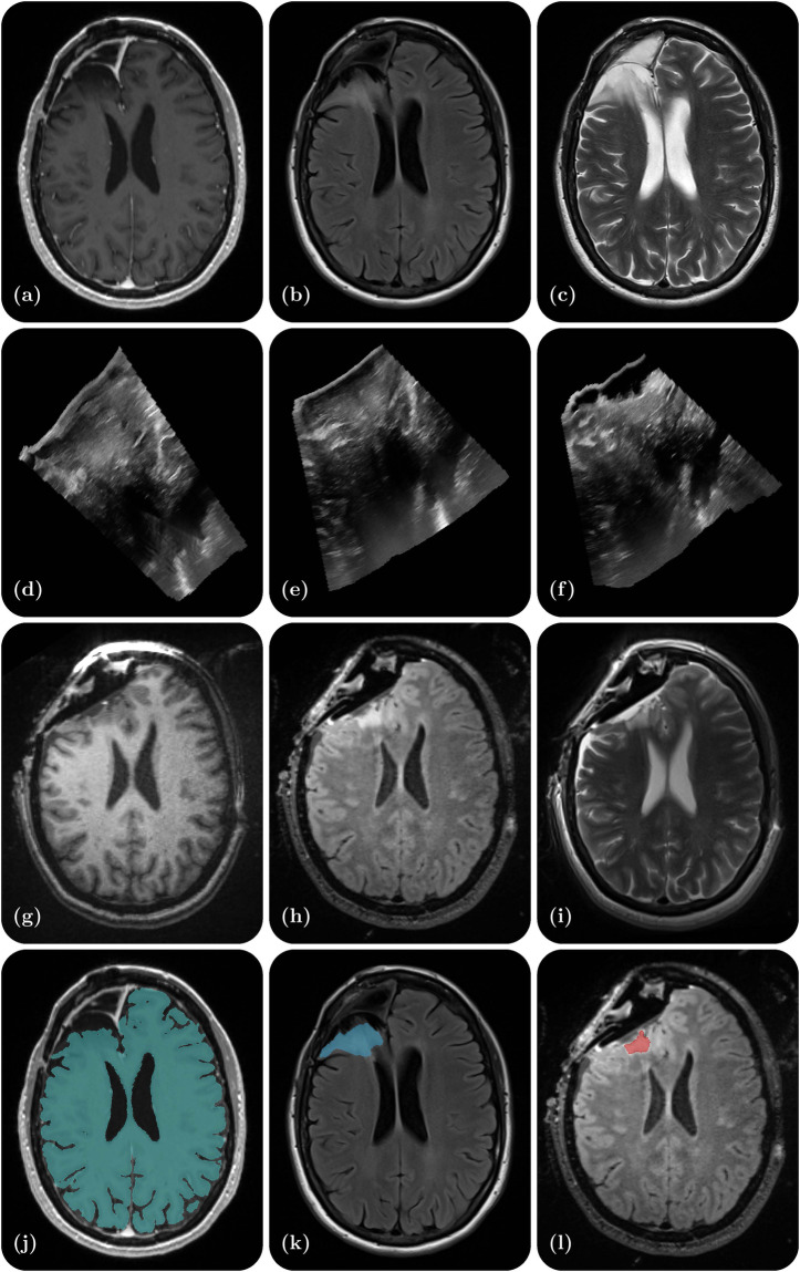
The standard of care for brain tumors is maximal safe surgical resection. Neuronavigation augments the surgeon's ability to achieve this but loses validity as surgery progresses due to brain shift. Moreover, gliomas are often indistinguishable from surrounding healthy brain tissue. Intraoperative magnetic resonance imaging (iMRI) and ultrasound (iUS) help visualize the tumor and brain shift. iUS is faster and easier to incorporate into surgical workflows but offers a lower contrast between tumorous and healthy tissues than iMRI. With the success of data-hungry Artificial Intelligence algorithms in medical image analysis, the benefits of sharing well-curated data cannot be overstated. To this end, we provide the largest publicly available MRI and iUS database of surgically treated brain tumors, including gliomas (n = 92), metastases (n = 11), and others (n = 11). This collection contains 369 preoperative MRI series, 320 3D iUS series, 301 iMRI series, and 356 segmentations collected from 114 consecutive patients at a single institution. This database is expected to help brain shift and image analysis research and neurosurgical training in interpreting iUS and iMRI.

摘要

脑肿瘤的治疗标准是最大限度地安全手术切除。神经导航增强了外科医生实现这一目标的能力,但由于脑移位,其有效性会逐渐丧失。此外,胶质瘤通常与周围健康脑组织难以区分。术中磁共振成像(iMRI)和超声(iUS)有助于观察肿瘤和脑移位。iUS 更快、更容易融入手术流程,但与 iMRI 相比,其在肿瘤和健康组织之间的对比度更低。随着数据密集型人工智能算法在医学图像分析中的成功,共享精心整理的数据的好处怎么强调都不为过。为此,我们提供了最大的公开可用于手术治疗脑肿瘤的 MRI 和 iUS 数据库,包括胶质瘤(n=92)、转移瘤(n=11)和其他肿瘤(n=11)。该数据库包含 369 个术前 MRI 系列、320 个 3D iUS 系列、301 个 iMRI 系列和 356 个从一家机构的 114 名连续患者中收集的分割。该数据库有望有助于脑移位和图像分析研究以及神经外科医生在解读 iUS 和 iMRI 方面的培训。

https://cdn.ncbi.nlm.nih.gov/pmc/blobs/aef0/11093985/4e8eae0a9803/41597_2024_3295_Fig1_HTML.jpg

相似文献

1
ReMIND: The Brain Resection Multimodal Imaging Database.ReMIND:脑切除多模态成像数据库。
Sci Data. 2024 May 14;11(1):494. doi: 10.1038/s41597-024-03295-z.
2
ReMIND: The Brain Resection Multimodal Imaging Database.ReMIND:脑切除多模态成像数据库。
medRxiv. 2024 Apr 8:2023.09.14.23295596. doi: 10.1101/2023.09.14.23295596.
3
Clinical application of 3.0 T intraoperative magnetic resonance combined with multimodal neuronavigation in resection of cerebral eloquent area glioma.3.0T术中磁共振联合多模态神经导航在脑功能区胶质瘤切除术中的临床应用
Medicine (Baltimore). 2018 Aug;97(34):e11702. doi: 10.1097/MD.0000000000011702.
4
Navigated intraoperative ultrasound in pediatric brain tumors.术中导航超声在小儿脑肿瘤中的应用。
Childs Nerv Syst. 2024 Sep;40(9):2697-2705. doi: 10.1007/s00381-024-06492-8. Epub 2024 Jun 11.
5
Challenges and Opportunities of Intraoperative 3D Ultrasound With Neuronavigation in Relation to Intraoperative MRI.术中三维超声与神经导航结合相对于术中磁共振成像的挑战与机遇
Front Oncol. 2021 May 3;11:656519. doi: 10.3389/fonc.2021.656519. eCollection 2021.
6
Factors triggering an additional resection and determining residual tumor volume on intraoperative MRI: analysis from a prospective single-center registry of supratentorial gliomas.触发额外切除并在术中磁共振成像上确定残余肿瘤体积的因素:来自幕上胶质瘤前瞻性单中心登记处的分析
Neurosurg Focus. 2016 Mar;40(3):E4. doi: 10.3171/2015.11.FOCUS15542.
7
[Application of intraoperative MRI combined with neuronavigation in microsurgical resection for 
insular glioma].术中磁共振成像联合神经导航在岛叶胶质瘤显微手术切除中的应用
Zhong Nan Da Xue Xue Bao Yi Xue Ban. 2018 Apr 28;43(4):383-387. doi: 10.11817/j.issn.1672-7347.2018.04.007.
8
Surgical benefits of combined awake craniotomy and intraoperative magnetic resonance imaging for gliomas associated with eloquent areas.联合清醒开颅术和术中磁共振成像在与功能区相关的脑胶质瘤中的手术获益。
J Neurosurg. 2017 Oct;127(4):790-797. doi: 10.3171/2016.9.JNS16152. Epub 2017 Jan 6.
9
Intraoperative Multi-Information-Guided Resection of Dominant-Sided Insular Gliomas in a 3-T Intraoperative Magnetic Resonance Imaging Integrated Neurosurgical Suite.在3T术中磁共振成像集成神经外科手术套件中对优势侧岛叶胶质瘤进行术中多信息引导下的切除术
World Neurosurg. 2016 May;89:84-92. doi: 10.1016/j.wneu.2016.01.067. Epub 2016 Feb 4.
10
Intraoperative Integration of Multimodal Imaging to Improve Neuronavigation: A Technical Note.术中多模态影像融合提高神经导航精度:一项技术说明。
World Neurosurg. 2022 Aug;164:330-340. doi: 10.1016/j.wneu.2022.05.133. Epub 2022 Jun 3.

引用本文的文献

1
A decentralized privacy-preserving XR system for 3D medical data visualization using hybrid biometric cryptosystem.一种使用混合生物特征密码系统的用于3D医学数据可视化的去中心化隐私保护XR系统。
Sci Rep. 2025 Aug 5;15(1):28568. doi: 10.1038/s41598-025-08784-8.
2
OMT and tensor SVD-based deep learning model for segmentation and predicting genetic markers of glioma: A multicenter study.基于OMT和张量奇异值分解的深度学习模型用于胶质瘤的分割和遗传标记预测:一项多中心研究。
Proc Natl Acad Sci U S A. 2025 Jul 15;122(28):e2500004122. doi: 10.1073/pnas.2500004122. Epub 2025 Jul 8.
3
Integrating artificial intelligence in healthcare: applications, challenges, and future directions.

本文引用的文献

1
Unified Brain MR-Ultrasound Synthesis using Multi-Modal Hierarchical Representations.使用多模态分层表示的统一脑磁共振-超声合成
Med Image Comput Comput Assist Interv. 2023 Oct 13;2023:448-458. doi: 10.1007/978-3-031-43999-5_43.
2
Challenges and Opportunities of Intraoperative 3D Ultrasound With Neuronavigation in Relation to Intraoperative MRI.术中三维超声与神经导航结合相对于术中磁共振成像的挑战与机遇
Front Oncol. 2021 May 3;11:656519. doi: 10.3389/fonc.2021.656519. eCollection 2021.
3
Learning joint segmentation of tissues and brain lesions from task-specific hetero-modal domain-shifted datasets.
将人工智能整合到医疗保健中:应用、挑战及未来方向。
Future Sci OA. 2025 Dec;11(1):2527505. doi: 10.1080/20565623.2025.2527505. Epub 2025 Jul 4.
4
Optimizing registration uncertainty visualization to support intraoperative decision-making during brain tumor resection.优化配准不确定性可视化以支持脑肿瘤切除术中的术中决策。
Int J Comput Assist Radiol Surg. 2025 May 13. doi: 10.1007/s11548-025-03407-z.
5
Triad: Vision Foundation Model for 3D Magnetic Resonance Imaging.三联征:用于三维磁共振成像的视觉基础模型
Res Sq. 2025 Mar 10:rs.3.rs-6129856. doi: 10.21203/rs.3.rs-6129856/v1.
6
Advancing Precision Oncology Through Modeling of Longitudinal and Multimodal Data.通过纵向和多模态数据建模推进精准肿瘤学
ArXiv. 2025 Apr 29:arXiv:2502.07836v2.
7
A database of magnetic resonance imaging-transcranial ultrasound co-registration.磁共振成像-经颅超声联合配准数据库。
Med Phys. 2025 May;52(5):3481-3486. doi: 10.1002/mp.17666. Epub 2025 Feb 7.
8
Exploring adult glioma through MRI: A review of publicly available datasets to guide efficient image analysis.通过磁共振成像探索成人胶质瘤:对公开可用数据集的综述以指导高效图像分析。
Neurooncol Adv. 2025 Jan 28;7(1):vdae197. doi: 10.1093/noajnl/vdae197. eCollection 2025 Jan-Dec.
9
Deep Learning-Based Glioma Segmentation of 2D Intraoperative Ultrasound Images: A Multicenter Study Using the Brain Tumor Intraoperative Ultrasound Database (BraTioUS).基于深度学习的二维术中超声图像胶质瘤分割:使用脑肿瘤术中超声数据库(BraTioUS)的多中心研究
Cancers (Basel). 2025 Jan 19;17(2):315. doi: 10.3390/cancers17020315.
10
DNA palette code for time-series archival data storage.用于时间序列存档数据存储的DNA调色板编码。
Natl Sci Rev. 2024 Sep 10;12(1):nwae321. doi: 10.1093/nsr/nwae321. eCollection 2025 Jan.
从特定任务的异模态域移位数据集学习组织和脑损伤的联合分割。
Med Image Anal. 2021 Jan;67:101862. doi: 10.1016/j.media.2020.101862. Epub 2020 Oct 9.
4
cIMPACT-NOW update 7: advancing the molecular classification of ependymal tumors.cIMPACT-NOW 更新 7:推进室管膜肿瘤的分子分类。
Brain Pathol. 2020 Sep;30(5):863-866. doi: 10.1111/bpa.12866. Epub 2020 Jun 23.
5
cIMPACT-NOW update 6: new entity and diagnostic principle recommendations of the cIMPACT-Utrecht meeting on future CNS tumor classification and grading.cIMPACT-NOW更新6:关于未来中枢神经系统肿瘤分类和分级的cIMPACT-乌得勒支会议的新实体和诊断原则建议。
Brain Pathol. 2020 Jul;30(4):844-856. doi: 10.1111/bpa.12832. Epub 2020 Apr 19.
6
Evaluation of MRI to Ultrasound Registration Methods for Brain Shift Correction: The CuRIOUS2018 Challenge.基于 MRI 与超声配准方法的脑移位校正评估:2018 年 CuRIOUS 挑战赛
IEEE Trans Med Imaging. 2020 Mar;39(3):777-786. doi: 10.1109/TMI.2019.2935060. Epub 2019 Aug 13.
7
cIMPACT-NOW update 4: diffuse gliomas characterized by MYB, MYBL1, or FGFR1 alterations or BRAF mutation.cIMPACT-NOW更新4:以MYB、MYBL1或FGFR1改变或BRAF突变为特征的弥漫性胶质瘤
Acta Neuropathol. 2019 Apr;137(4):683-687. doi: 10.1007/s00401-019-01987-0. Epub 2019 Mar 8.
8
SimpleITK Image-Analysis Notebooks: a Collaborative Environment for Education and Reproducible Research.SimpleITK 图像分析笔记本:用于教育和可重复研究的协作环境。
J Digit Imaging. 2018 Jun;31(3):290-303. doi: 10.1007/s10278-017-0037-8.
9
: An Open Source Library for Standardized Communication of Quantitative Image Analysis Results Using DICOM.一个用于使用DICOM进行定量图像分析结果标准化通信的开源库。
Cancer Res. 2017 Nov 1;77(21):e87-e90. doi: 10.1158/0008-5472.CAN-17-0336.
10
WHO 2016 Classification of gliomas.世界卫生组织 2016 年版《脑肿瘤分类》。
Neuropathol Appl Neurobiol. 2018 Feb;44(2):139-150. doi: 10.1111/nan.12432.