Song Joo Hye, Kim Eun Ran, Hong Yiyu, Sohn Insuk, Ahn Soomin, Kim Seok-Hyung, Jang Kee-Taek
Department of Internal Medicine, Konkuk University Medical Center, Konkuk University School of Medicine, Seoul 05030, Republic of Korea.
Department of Medicine, Samsung Medical Center, Sungkyunkwan University School of Medicine, Seoul 06351, Republic of Korea.
Cancers (Basel). 2024 May 16;16(10):1900. doi: 10.3390/cancers16101900.
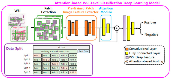
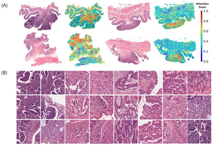
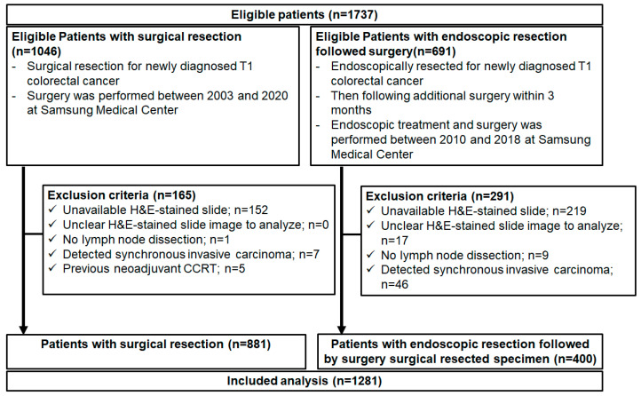
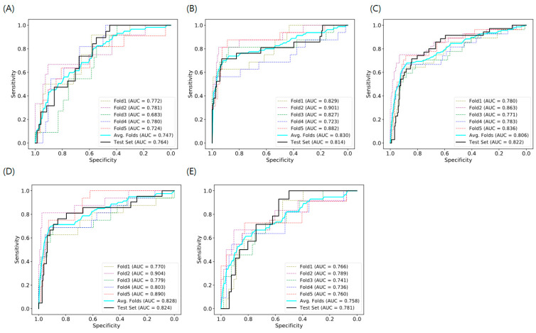
According to the current guidelines, additional surgery is performed for endoscopically resected specimens of early colorectal cancer (CRC) with a high risk of lymph node metastasis (LNM). However, the rate of LNM is 2.1-25.0% in cases treated endoscopically followed by surgery, indicating a high rate of unnecessary surgeries. Therefore, this study aimed to develop an artificial intelligence (AI) model using H&E-stained whole slide images (WSIs) without handcrafted features employing surgically and endoscopically resected specimens to predict LNM in T1 CRC. To validate with an independent cohort, we developed a model with four versions comprising various combinations of training and test sets using H&E-stained WSIs from endoscopically (400 patients) and surgically resected specimens (881 patients): Version 1, Train and Test: surgical specimens; Version 2, Train and Test: endoscopic and surgically resected specimens; Version 3, Train: endoscopic and surgical specimens and Test: surgical specimens; Version 4, Train: endoscopic and surgical specimens and Test: endoscopic specimens. The area under the curve (AUC) of the receiver operating characteristic curve was used to determine the accuracy of the AI model for predicting LNM with a 5-fold cross-validation in the training set. Our AI model with H&E-stained WSIs and without annotations showed good performance power with the validation of an independent cohort in a single center. The AUC of our model was 0.758-0.830 in the training set and 0.781-0.824 in the test set, higher than that of previous AI studies with only WSI. Moreover, the AI model with Version 4, which showed the highest sensitivity (92.9%), reduced unnecessary additional surgery by 14.2% more than using the current guidelines (68.3% vs. 82.5%). This revealed the feasibility of using an AI model with only H&E-stained WSIs to predict LNM in T1 CRC.
根据当前指南,对于内镜切除的早期结直肠癌(CRC)标本且具有高淋巴结转移(LNM)风险的患者需进行额外手术。然而,在内镜治疗后再行手术的病例中,LNM发生率为2.1%-25.0%,这表明不必要手术的比例很高。因此,本研究旨在开发一种人工智能(AI)模型,该模型使用苏木精-伊红(H&E)染色的全切片图像(WSIs),无需手工特征,利用手术切除和内镜切除的标本预测T1期CRC中的LNM。为了用独立队列进行验证,我们开发了一个包含四个版本的模型,这些版本使用来自内镜切除标本(400例患者)和手术切除标本(881例患者)的H&E染色WSIs,包括训练集和测试集的各种组合:版本1,训练和测试:手术标本;版本2,训练和测试:内镜和手术切除标本;版本3,训练:内镜和手术标本,测试:手术标本;版本4,训练:内镜和手术标本,测试:内镜标本。在训练集中,采用5折交叉验证,使用受试者工作特征曲线下面积(AUC)来确定AI模型预测LNM的准确性。我们的带有H&E染色WSIs且无注释的AI模型在单中心独立队列验证中表现出良好的性能。我们模型在训练集中的AUC为0.758-0.830,在测试集中为0.781-0.824,高于以往仅使用WSIs的AI研究。此外,版本4的AI模型显示出最高的灵敏度(92.9%),比使用当前指南减少了14.2%的不必要额外手术(68.3%对82.5%)。这揭示了仅使用H&E染色WSIs的AI模型预测T1期CRC中LNM的可行性。