Department of Ophthalmology, Hospital Universitari de Bellvitge, Carrer de La Feixa Llarga, S/N, 08907, L'Hospitalet de Llobregat, Catalunya, Spain.
Institut de La Màcula, 08022, Barcelona, Spain.
Acta Diabetol. 2024 Nov;61(11):1385-1392. doi: 10.1007/s00592-024-02290-5. Epub 2024 May 27.
To determine the presence of sectoral changes in vessel density (VD) patterns induced by vascular endothelial growth factor inhibitors (anti-VEGF) in patients with diabetic macular edema (DME) using optical coherence tomography angiography (OCTA).
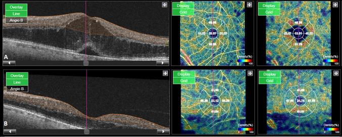
Prospective, interventional study. A total of 43 patients (63 eyes) were initially enrolled in the study. We performed swept source (SS) OCT and sectorial OCTA measurement to determine parafoveal VD at baseline and after six months of anti-VEGF treatment. In the locations with statistically significant differences in VD between baseline and month 6, we performed univariate and multivariate analyses to determine which, if any, of the baseline variables were associated with the observed changes.
A total of 34 patients (48 eyes) were included in the final analysis. Mean VD decreased from baseline to month 6 (from 45.2 (± 3.5) to 44.6 (± 3.2) % in the SCP and from 50 (± 3.3) to 49 (± 3.9) % in the DCP). The only significant changes in VD were observed in the nasal sector of the deep capillary plexus, with a decrease of 2.9% (p = 0.001). On univariate and multivariate analyses, the only variable significantly associated with changes in VD in the nasal sector after 6 months of treatment was baseline VD in the same sector.
Anti-VEGF therapy has a small impact on VD values over time. These variations observed after treatment seems to be related to changes over areas of vascular anomalies and displaced vessels adjacent to cystic areas, with no significant changes over ischemic areas. No correlation was observed between this trend and other clinical baseline features.
使用光相干断层扫描血管造影术(OCTA)确定血管内皮生长因子抑制剂(抗 VEGF)治疗糖尿病黄斑水肿(DME)患者血管密度(VD)模式的扇形变化。
前瞻性、干预性研究。最初有 43 名患者(63 只眼)入组该研究。我们进行了扫频源(SS)OCT 和扇形 OCTA 测量,以确定基线时和抗 VEGF 治疗 6 个月后的中心凹旁 VD。在 VD 基线和 6 个月时存在统计学差异的位置,我们进行了单变量和多变量分析,以确定观察到的变化与任何基线变量相关。
共有 34 名患者(48 只眼)纳入最终分析。平均 VD 从基线到 6 个月时下降(SCP 从 45.2(±3.5)%降至 44.6(±3.2)%,DCP 从 50(±3.3)%降至 49(±3.9)%)。仅在深层毛细血管丛的鼻侧扇形区观察到 VD 有显著变化,下降了 2.9%(p=0.001)。在单变量和多变量分析中,治疗 6 个月后鼻侧 VD 变化与唯一显著相关的变量是同一部位的基线 VD。
抗 VEGF 治疗随时间推移对 VD 值的影响较小。治疗后观察到的这些变化似乎与血管异常和邻近囊腔的移位血管区域的变化有关,与缺血区域无显著变化。这种趋势与其他临床基线特征之间没有相关性。