Klangprapan Jutapak, Souza Glauco R, Ferreira João N
Avatar Biotechnologies for Oral Health and Healthy Longevity Research Unit, Faculty of Dentistry, Chulalongkorn University, 34 Henri-Dunant Road, Pathumwan, Bangkok, 10330, Thailand.
Greiner Bio-one North America Inc., 4238 Capital Drive, Monroe, NC, 28110, USA.
BDJ Open. 2024 May 30;10(1):39. doi: 10.1038/s41405-024-00219-2.
Salivary gland (SG) hypofunction is a common clinical condition arising from radiotherapy to suppress head and neck cancers. The radiation often destroys the SG secretory acini, and glands are left with limited regenerative potential. Due to the complex architecture of SG acini and ducts, three-dimensional (3D) bioprinting platforms have emerged to spatially define these in vitro epithelial units and develop mini-organs or organoids for regeneration. Due to the limited body of evidence, this comprehensive review highlights the advantages and challenges of bioprinting platforms for SG regeneration.
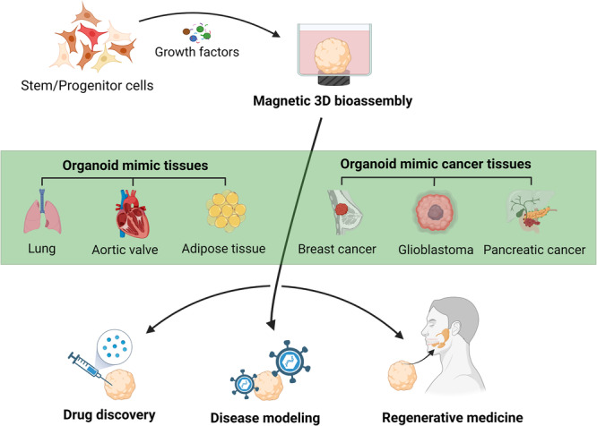
SG microtissue engineering strategies such as magnetic 3D bioassembly of cells and microfluidic coaxial 3D bioprinting of cell-laden microfibers and microtubes have been proposed to replace the damaged acinar units, avoid the use of xenogeneic matrices (like Matrigel), and restore salivary flow.
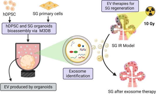
Replacing the SG damaged organ is challenging due to its complex architecture, which combines a ductal network with acinar epithelial units to facilitate a unidirectional flow of saliva. Our research group was the first to develop 3D bioassembly SG epithelial functional organoids with innervation to respond to both cholinergic and adrenergic stimulation. More recently, microtissue engineering using coaxial 3D bioprinting of hydrogel microfibers and microtubes could also supported the formation of viable epithelial units. Both bioprinting approaches could overcome the need for Matrigel by facilitating the assembly of adult stem cells, such as human dental pulp stem cells, and primary SG cells into micro-sized 3D constructs able to produce their own matrix and self-organize into micro-modular tissue clusters with lumenized areas. Furthermore, extracellular vesicle (EV) therapies from organoid-derived secretome were also designed and validated ex vivo for SG regeneration after radiation damage.
Magnetic 3D bioassembly and microfluidic coaxial bioprinting platforms have the potential to create SG mini-organs for regenerative applications via organoid transplantation or organoid-derived EV therapies.
唾液腺功能减退是头颈部癌症放射治疗引发的一种常见临床病症。放疗常破坏唾液腺分泌腺泡,且腺体的再生潜力有限。由于唾液腺腺泡和导管的结构复杂,三维(3D)生物打印平台应运而生,用于在空间上界定这些体外上皮单元,并开发用于再生的微型器官或类器官。鉴于证据有限,本综述重点介绍生物打印平台在唾液腺再生方面的优势和挑战。
已提出唾液腺微组织工程策略,如细胞的磁性3D生物组装以及载细胞微纤维和微管的微流控同轴3D生物打印,以替代受损的腺泡单元,避免使用异种基质(如基质胶),并恢复唾液分泌。
由于唾液腺结构复杂,其导管网络与腺泡上皮单元相结合以促进唾液单向流动,因此替换受损的唾液腺器官具有挑战性。我们的研究小组率先开发出具有神经支配的3D生物组装唾液腺上皮功能类器官,以响应胆碱能和肾上腺素能刺激。最近,使用水凝胶微纤维和微管的同轴3D生物打印进行微组织工程也能够支持有活力的上皮单元的形成。两种生物打印方法都可以通过促进成体干细胞(如人牙髓干细胞)和原代唾液腺细胞组装成能够产生自身基质并自组织成具有腔化区域的微型模块化组织簇的微型3D构建体,从而避免使用基质胶。此外,还设计并在体外验证了源自类器官分泌组的细胞外囊泡(EV)疗法对辐射损伤后唾液腺再生的作用。
磁性3D生物组装和微流控同轴生物打印平台有潜力通过类器官移植或类器官衍生的EV疗法创建用于再生应用的唾液腺微型器官。