STADIUS Center for Dynamical Systems, Signal Processing, and Data Analytics, Department of Electrical Engineering (ESAT), KU Leuven, Leuven, Belgium.
Aspect Analytics NV, Genk, Belgium.
PLoS One. 2024 May 31;19(5):e0304709. doi: 10.1371/journal.pone.0304709. eCollection 2024.
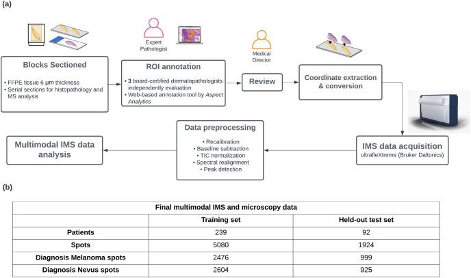
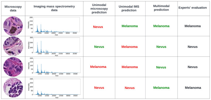
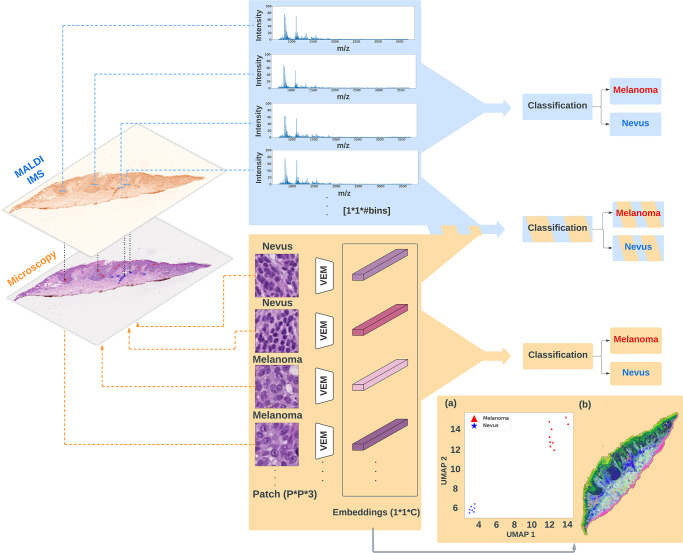
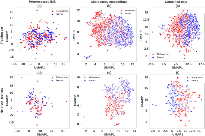
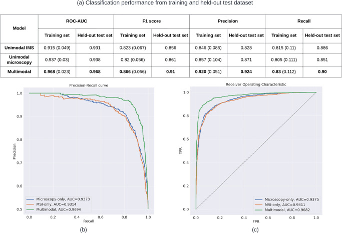
Imaging mass spectrometry (IMS) provides promising avenues to augment histopathological investigation with rich spatio-molecular information. We have previously developed a classification model to differentiate melanoma from nevi lesions based on IMS protein data, a task that is challenging solely by histopathologic evaluation. Most IMS-focused studies collect microscopy in tandem with IMS data, but this microscopy data is generally omitted in downstream data analysis. Microscopy, nevertheless, forms the basis for traditional histopathology and thus contains invaluable morphological information. In this work, we developed a multimodal classification pipeline that uses deep learning, in the form of a pre-trained artificial neural network, to extract the meaningful morphological features from histopathological images, and combine it with the IMS data. To test whether this deep learning-based classification strategy can improve on our previous results in classification of melanocytic neoplasia, we utilized MALDI IMS data with collected serial H&E stained sections for 331 patients, and compared this multimodal classification pipeline to classifiers using either exclusively microscopy or IMS data. The multimodal pipeline achieved the best performance, with ROC-AUCs of 0.968 vs. 0.938 vs. 0.931 for the multimodal, unimodal microscopy and unimodal IMS pipelines respectively. Due to the use of a pre-trained network to perform the morphological feature extraction, this pipeline does not require any training on large amounts of microscopy data. As such, this framework can be readily applied to improve classification performance in other experimental settings where microscopy data is acquired in tandem with IMS experiments.
成像质谱 (IMS) 提供了有前途的途径,通过丰富的空间 - 分子信息来增强组织病理学研究。我们之前开发了一种分类模型,基于 IMS 蛋白质数据来区分黑色素瘤和痣病变,这是仅通过组织病理学评估具有挑战性的任务。大多数专注于 IMS 的研究都与 IMS 数据一起收集显微镜图像,但这些显微镜数据通常在下游数据分析中被省略。然而,显微镜构成了传统组织病理学的基础,因此包含了宝贵的形态学信息。在这项工作中,我们开发了一种多模态分类管道,该管道使用深度学习,以预先训练的人工神经网络的形式,从组织病理学图像中提取有意义的形态特征,并将其与 IMS 数据相结合。为了测试这种基于深度学习的分类策略是否可以改进我们之前在黑色素瘤分类中的结果,我们利用 MALDI IMS 数据和收集的 331 名患者的连续 H&E 染色切片进行测试,并将这种多模态分类管道与仅使用显微镜或 IMS 数据的分类器进行比较。多模态管道的性能最佳,多模态、单模态显微镜和单模态 IMS 管道的 ROC-AUC 分别为 0.968、0.938 和 0.931。由于使用预先训练的网络来执行形态特征提取,因此该管道不需要在大量显微镜数据上进行任何训练。因此,这种框架可以很容易地应用于其他实验设置中,在这些实验设置中,显微镜数据与 IMS 实验同时采集,以提高分类性能。