Department of Dermatology, Erasmus University Medical Center, Rotterdam, The Netherlands.
Department of Plastic, Reconstructive, and Aesthetic Surgery, N2 Aesthetics, Manhattan Beach, CA.
Dermatol Surg. 2024 Oct 1;50(10):946-952. doi: 10.1097/DSS.0000000000004257. Epub 2024 Jun 4.
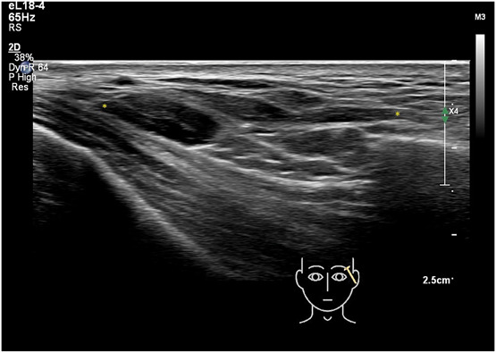
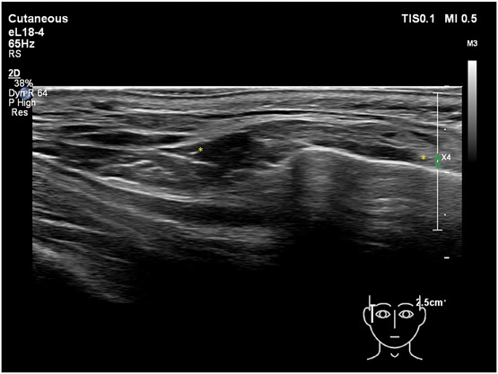
Clinical and ultrasound experience has revealed that after soft tissue injections of the lateral cheek, the filler may displace from the zygoma to the caudal temporal area.
To obtain more data to provide insight into product distribution when soft tissue fillers are injected in the zygomatic region.
Two hundred patients were examined with facial ultrasound imaging of the zygomatic and temporal region. Inclusion criteria were simply a positive response on the screening questionnaire as to whether or not they had filler injections placed in their lateral cheek. Control injections were also performed to the zygomatic regions of a body donor and in 10 patients ultrasound-guided.
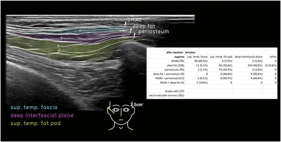
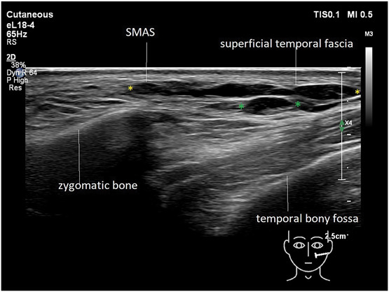
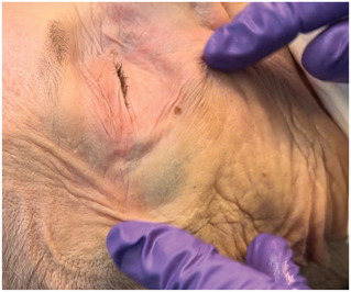
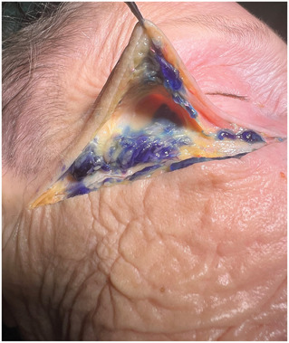
A correlation was found between the layers in which filler was detected on the zygoma and where it was ultimately found in the temples. Four different redistribution patterns were observed: (1) migration of filler within the superficial muscular aponeurotic system (SMAS) on the zygoma into the superficial temporal fascia. Migration of filler from the lateral suborbicularis oculi fat to (2) the deep interfacial plane of the temple or (3) to the superficial temporal fat pad; (4) migration from the supraperiosteal layer of the zygoma to the superficial temporal fat pad. Body donor and patients: filler deposits injected on the zygoma were witnessed to shift during injection into the caudal part of the temple.
Soft tissue filler aliquots may be redistributed into the temples after injections of the lateral side of the zygomatic arch. The displacement follows a distinct pattern depending on the initial layer of injection.
临床和超声经验表明,在面颊软组织注射后,填充物可能会从颧骨移位到颞区尾部。
获得更多数据,深入了解在颧骨区域注射软组织填充物时的产品分布情况。
对 200 名患者进行了颧骨和颞区的面部超声成像检查。纳入标准是在筛查问卷中简单地回答是否在外侧脸颊注射了填充物。还对尸体捐赠者的颧骨区域和 10 名患者进行了超声引导的对照注射。
在颧骨上检测到填充物的层次与在颞部最终发现填充物的位置之间存在相关性。观察到四种不同的再分布模式:(1)在颧骨的浅层肌肉腱膜系统(SMAS)内的填充物迁移到颞浅筋膜。填充物从外侧眼轮匝肌脂肪向(2)颞深界面或(3)颞浅脂肪垫迁移;(4)从颧骨的骨膜下层迁移到颞浅脂肪垫。尸体捐赠者和患者:在颧骨上注射的填充物在注射过程中被观察到移位到颞部尾部。
在颧骨侧注射后,软组织填充物可能会重新分布到颞部。这种移位遵循一个明确的模式,取决于注射的初始层。