Hacettepe University Faculty of Medicine, Department of Radiology, Division of Pediatric Radiology, Ankara, Türkiye
Hacettepe University Faculty of Medicine, Department of Radiology, Ankara, Türkiye
Diagn Interv Radiol. 2024 Nov 6;30(6):419-426. doi: 10.4274/dir.2024.242714. Epub 2024 Jun 10.
Pediatric lung tumors are primarily discussed in the surgical literature. However, limited research has been reported on their imaging findings, and only a few tumor types have been documented. Therefore, the aim of this article is to describe the imaging features of primary lung tumors in children.
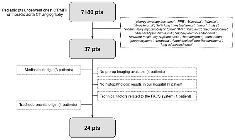
The archives of the pediatric radiology unit were reviewed for primary lung tumors documented between 2007 and 2023. In total, 24 patients (9 girls and 15 boys; aged 5 months to 16 years) were included in the study. Their demographic characteristics, clinical presentation, and histopathologic results were obtained. All imaging studies were reviewed by two radiologists for various findings (e.g., lymphadenopathy, atelectasis, pleural effusion, calcification, multiplicity, pneumothorax, axial and lobar location, laterality, tumor margin, mediastinal shift, contrast enhancement pattern, signal intensity on T1- and T2-weighted images, and diffusion pattern), and a final decision was made by consensus. The mean tumor size was compared between the benign and malignant groups using a t-test.
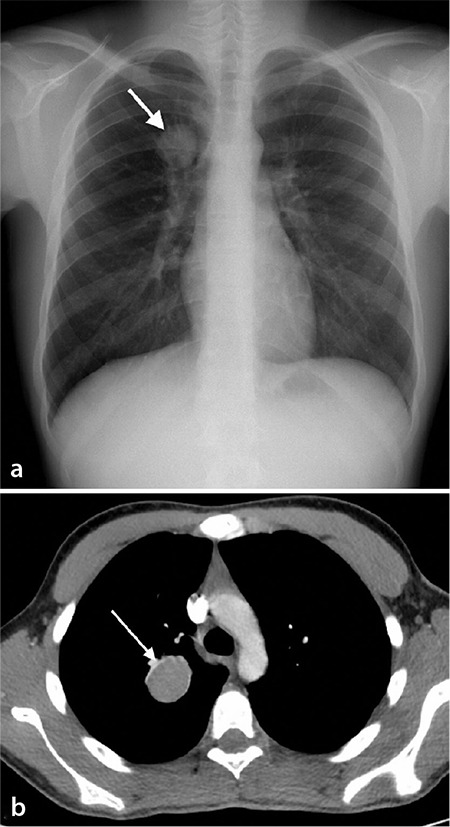
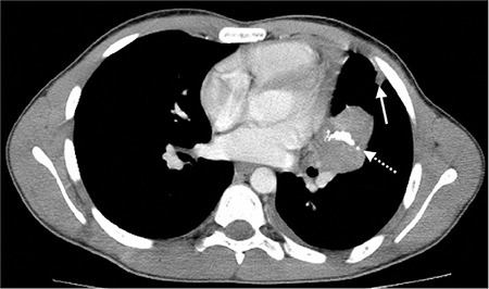
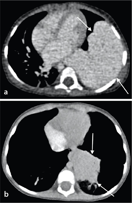
There were 15 (62.5%) benign tumors, as follows: inflammatory myofibroblastic tumor (IMT; n = 10, 41%), hemangioma (n = 2, 8%), pneumocytoma (n = 2, 8%), and mature cystic teratoma (n = 1, 4%). Moreover, there were 9 (37.5%) malignant tumors, as follows: pleuropulmonary blastoma (PPB; n = 6, 25%), adenocarcinoma (n = 2, 8%), and lymphoepithelioma-like carcinoma (LELC) (n = 1, 4%). The most frequently reported symptoms were cough, fever, dyspnea, chest pain, and recurrent infection; six patients reported no clinical symptoms. Fifteen tumors (62%) were located in the right lung. The mean tumor diameter at the time of diagnosis was 6.4 ± 3 cm (benign group: 6.7 ± 3.4 cm; malignant group: 6 ± 2.3 cm, > 0.050). Calcification was present in 80% of the patients with IMT. At the time of diagnosis, two (8.3%) patients were found to have metastasis: one was diagnosed with adenocarcinoma and the other with LELC. Tumors were located peripherally in 18 (75%) patients.
The symptoms associated with lung masses are non-specific. There is no correlation between tumor size and malignancy. The most common tumors observed in this study were IMT and PPB, respectively. IMT is highly associated with calcification.
Primary lung tumors are rarely seen in children, and they have different histopathological types. Calcification might be an important radiological clue for the diagnosis of IMT, which is the most common lung tumor in children.
小儿肺肿瘤主要在外科文献中讨论。然而,关于其影像学表现的研究报道有限,且仅记录了少数几种肿瘤类型。因此,本文旨在描述儿童原发性肺肿瘤的影像学特征。
回顾 2007 年至 2023 年间在小儿放射科记录的原发性肺肿瘤病例。共有 24 名患者(9 名女孩和 15 名男孩;年龄 5 个月至 16 岁)纳入研究。获取他们的人口统计学特征、临床表现和组织病理学结果。由两名放射科医生对所有影像学研究进行回顾,以评估各种发现(例如,淋巴结病、肺不张、胸腔积液、钙化、多发性、气胸、轴向和肺叶位置、侧别、肿瘤边缘、纵隔移位、对比增强模式、T1 和 T2 加权图像上的信号强度以及扩散模式),并通过共识做出最终决定。使用 t 检验比较良性和恶性组的平均肿瘤大小。
有 15 例(62.5%)为良性肿瘤,如下:炎症性肌纤维母细胞瘤(IMT;n = 10,41%)、血管瘤(n = 2,8%)、肺母细胞瘤(n = 2,8%)和成熟囊性畸胎瘤(n = 1,4%)。此外,有 9 例(37.5%)为恶性肿瘤,如下:胸膜肺母细胞瘤(PPB;n = 6,25%)、腺癌(n = 2,8%)和淋巴上皮样癌(LELC;n = 1,4%)。最常报告的症状是咳嗽、发热、呼吸困难、胸痛和反复感染;有 6 名患者无症状。15 个肿瘤(62%)位于右肺。诊断时的平均肿瘤直径为 6.4 ± 3 cm(良性组:6.7 ± 3.4 cm;恶性组:6 ± 2.3 cm,>0.050)。80%的 IMT 患者存在钙化。在诊断时,有 2 例(8.3%)患者发现转移:1 例诊断为腺癌,另 1 例诊断为 LELC。18 例(75%)患者的肿瘤位于外周。
与肺部肿块相关的症状是非特异性的。肿瘤大小与恶性程度之间无相关性。本研究中最常见的肿瘤是 IMT 和 PPB。IMT 与钙化密切相关。
儿童原发性肺肿瘤罕见,具有不同的组织病理学类型。钙化可能是诊断 IMT 的重要影像学线索,IMT 是儿童最常见的肺部肿瘤。