• 文献检索
  • 文档翻译
  • 深度研究
  • 学术资讯
  • Suppr Zotero 插件Zotero 插件
  • 邀请有礼
  • 套餐&价格
  • 历史记录
应用&插件
Suppr Zotero 插件Zotero 插件浏览器插件Mac 客户端Windows 客户端微信小程序
定价
高级版会员购买积分包购买API积分包
服务
文献检索文档翻译深度研究API 文档MCP 服务
关于我们
关于 Suppr公司介绍联系我们用户协议隐私条款
关注我们

Suppr 超能文献

核心技术专利:CN118964589B侵权必究
粤ICP备2023148730 号-1Suppr @ 2026

文献检索

告别复杂PubMed语法,用中文像聊天一样搜索,搜遍4000万医学文献。AI智能推荐,让科研检索更轻松。

立即免费搜索

文件翻译

保留排版,准确专业,支持PDF/Word/PPT等文件格式,支持 12+语言互译。

免费翻译文档

深度研究

AI帮你快速写综述,25分钟生成高质量综述,智能提取关键信息,辅助科研写作。

立即免费体验

实时成像辅助下为门静脉分叉缺如患者行腹腔镜左肝切除术:1例病例报告

Laparoscopic left hepatectomy for a patient with an absence of portal bifurcation using real-time imaging: a case report.

作者信息

Mizuno Shugo, Iizawa Yusuke, Tanemura Akihiro, Kaluba Benson, Noguchi Daisuke, Ito Takahiro, Hayasaki Aoi, Fujii Takehiro, Murata Yasuhiro, Kuriyama Naohisa, Kishiwada Masashi

机构信息

Department of Hepatobiliary Pancreatic and Transplant Surgery, Mie University School of Medicine, 2-174 Edobashi, Tsu, Mie, 514-8507, Japan.

出版信息

Surg Case Rep. 2024 Jun 11;10(1):140. doi: 10.1186/s40792-024-01945-3.

DOI:10.1186/s40792-024-01945-3
PMID:38861107
原文链接:https://pmc.ncbi.nlm.nih.gov/articles/PMC11166617/
Abstract

BACKGROUND

Absence of portal bifurcation is an extremely rare anomaly that should be recognized preoperatively, especially prior to a major hepatectomy.

CASE PRESENTATION

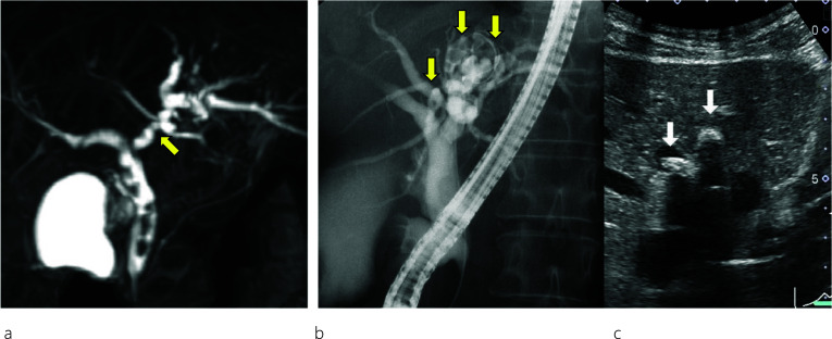
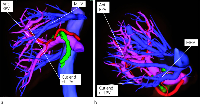
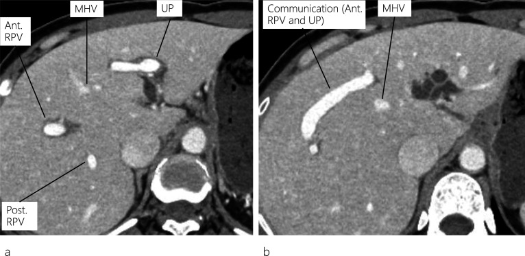
A 45-year-old woman presented with abdominal pain, and abdominal computed tomography (CT) revealed dilatation of both the common bile duct (CBD) and intrahepatic bile duct (IHBD). Endoscopic retrograde cholangiopancreatography (ERCP) showed CBD and IHBD stones (B2 and B4). The CBD stones were removed, but the IHBD stones could not be, yet there was no evidence of malignancy at the site of IHBD stenosis. Enhanced CT revealed a dilated IHBD, while three-dimensional CT images showed the left portal vein running through the ventral side of the middle hepatic vein, which was diagnosed as the absence of portal vein bifurcation (APB). Laparoscopic left hepatectomy was successfully performed using real-time indocyanine green (ICG) fluorescence imaging.

CONCLUSION

Surgeons should be aware of the possibility of APB, a rare portal vein anomaly, before performing major hepatectomy. Real-time ICG fluorescence imaging may be helpful to ensure the precise anatomy of the liver during laparoscopic surgery.

摘要

背景

门静脉分支缺如是一种极其罕见的异常情况,应在术前,尤其是在进行大型肝切除术前予以识别。

病例报告

一名45岁女性因腹痛就诊,腹部计算机断层扫描(CT)显示胆总管(CBD)和肝内胆管(IHBD)均扩张。内镜逆行胰胆管造影(ERCP)显示CBD和IHBD结石(B2和B4)。CBD结石已取出,但IHBD结石未能取出,且IHBD狭窄部位无恶性肿瘤迹象。增强CT显示IHBD扩张,而三维CT图像显示左门静脉穿过肝中静脉腹侧,诊断为门静脉分支缺如(APB)。使用实时吲哚菁绿(ICG)荧光成像成功实施了腹腔镜左肝切除术。

结论

外科医生在进行大型肝切除术前应意识到APB这种罕见门静脉异常情况的可能性。实时ICG荧光成像可能有助于在腹腔镜手术期间确保肝脏的精确解剖。

https://cdn.ncbi.nlm.nih.gov/pmc/blobs/29ea/11166617/1f4b79e052f5/40792_2024_1945_Fig5_HTML.jpg
https://cdn.ncbi.nlm.nih.gov/pmc/blobs/29ea/11166617/21e96c4818fd/40792_2024_1945_Fig1_HTML.jpg
https://cdn.ncbi.nlm.nih.gov/pmc/blobs/29ea/11166617/69e936b05edd/40792_2024_1945_Fig2_HTML.jpg
https://cdn.ncbi.nlm.nih.gov/pmc/blobs/29ea/11166617/4715a31e6abe/40792_2024_1945_Fig3_HTML.jpg
https://cdn.ncbi.nlm.nih.gov/pmc/blobs/29ea/11166617/0c56e9d234af/40792_2024_1945_Fig4_HTML.jpg
https://cdn.ncbi.nlm.nih.gov/pmc/blobs/29ea/11166617/1f4b79e052f5/40792_2024_1945_Fig5_HTML.jpg

相似文献

1
Laparoscopic left hepatectomy for a patient with an absence of portal bifurcation using real-time imaging: a case report.实时成像辅助下为门静脉分叉缺如患者行腹腔镜左肝切除术:1例病例报告
Surg Case Rep. 2024 Jun 11;10(1):140. doi: 10.1186/s40792-024-01945-3.
2
Laparoscopic ultrasound-guided superselective portal vein injection combined with real-time indocyanine green fluorescence imaging and navigation for accurate resection of localized intrahepatic bile duct dilatation: a case report.腹腔镜超声引导下超选择性门静脉注射联合实时吲哚菁绿荧光成像和导航用于精准切除局限性肝内胆管扩张:一例报告。
BMC Surg. 2021 Aug 17;21(1):328. doi: 10.1186/s12893-021-01325-w.
3
Video-Based Indocyanine Green Fluorescence Applied to Robotic Duodenum-Preserving Pancreatic Head Resection.基于视频的吲哚菁绿荧光在机器人保留十二指肠胰头切除术的应用。
Ann Surg Oncol. 2024 Apr;31(4):2654-2655. doi: 10.1245/s10434-024-14911-y. Epub 2024 Jan 25.
4
Robotic Klatskin Type 3A Resection with Biliary Reconstruction: Description of Surgical Technique and Outcomes of Initial Series.机器人 Klatskin 型 3A 切除术联合胆道重建:手术技术描述及初步系列结果。
Ann Surg Oncol. 2023 Dec;30(13):8559-8560. doi: 10.1245/s10434-023-14256-y. Epub 2023 Sep 8.
5
Indocyanine Green-Enhanced Fluorescence in Laparoscopic Duodenum-Preserving Pancreatic Head Resection: Technique with Video.腹腔镜保留十二指肠胰头切除术的吲哚菁绿增强荧光:附有视频的技术。
Ann Surg Oncol. 2020 Oct;27(10):3926-3927. doi: 10.1245/s10434-020-08360-6. Epub 2020 Apr 7.
6
Pure Laparoscopic Living Donor Right Hepatectomy Using Real-Time Indocyanine Green Fluorescence Imaging.实时吲哚菁绿荧光成像引导下的纯腹腔镜活体右半肝切除术。
J Gastrointest Surg. 2019 Aug;23(8):1711-1712. doi: 10.1007/s11605-019-04217-w. Epub 2019 May 31.
7
Resection of a hilar cholangiocarcinoma in a patient with absent portal bifurcation.门静脉分叉缺如患者肝门部胆管癌切除术
Hepatogastroenterology. 2000 Mar-Apr;47(32):362-4.
8
Portal vein injury following endoscopic retrograde cholangiopancreatography: A case report.内镜逆行胰胆管造影术后门静脉损伤:1 例报告。
Ulus Travma Acil Cerrahi Derg. 2023 Mar;29(3):443-447. doi: 10.14744/tjtes.2022.28923.
9
Description of the Vascular Anatomy of Livers with Absence of the Portal Bifurcation.无门静脉分叉肝脏的血管解剖描述。
World J Surg. 2021 Mar;45(3):833-840. doi: 10.1007/s00268-020-05848-w. Epub 2020 Nov 9.
10
Laparoscopic Anatomical Portal Territory Hepatectomy with Cirrhosis by Takasaki's Approach and Indocyanine Green Fluorescence Navigation (with Video).经 Takasaki 入路的腹腔镜解剖性门脉蒂肝切除术治疗肝硬化合并症:吲哚菁绿荧光导航技术(附视频)。
Ann Surg Oncol. 2020 Dec;27(13):5179-5180. doi: 10.1245/s10434-020-08592-6. Epub 2020 May 19.

本文引用的文献

1
Description of the Vascular Anatomy of Livers with Absence of the Portal Bifurcation.无门静脉分叉肝脏的血管解剖描述。
World J Surg. 2021 Mar;45(3):833-840. doi: 10.1007/s00268-020-05848-w. Epub 2020 Nov 9.
2
Indocyanine green fluorescence imaging techniques and interventional radiology during laparoscopic anatomical liver resection (with video).吲哚菁绿荧光成像技术与介入放射学在腹腔镜解剖性肝切除术中的应用(附视频)。
Surg Endosc. 2018 Feb;32(2):1051-1055. doi: 10.1007/s00464-017-5997-8. Epub 2017 Dec 22.
3
Applications of fusion-fluorescence imaging using indocyanine green in laparoscopic hepatectomy.
融合荧光成像技术在腹腔镜肝切除术中应用吲哚菁绿的研究进展。
Surg Endosc. 2017 Dec;31(12):5111-5118. doi: 10.1007/s00464-017-5576-z. Epub 2017 Apr 28.
4
Complication of portal vein thrombosis after right hemihepatectomy in a patient lacking the portal vein bifurcation.门静脉分支缺如患者右半肝切除术后门静脉血栓形成并发症
J Med Invest. 2016;63(3-4):315-8. doi: 10.2152/jmi.63.315.
5
Right hemihepatectomy in presence of congenital absence of portal vein bifurcation: a challenging but feasible procedure.先天性门静脉分支缺如情况下的右半肝切除术:一项具有挑战性但可行的手术。
J Am Coll Surg. 2012 Aug;215(2):e1-4. doi: 10.1016/j.jamcollsurg.2012.05.016. Epub 2012 Jun 8.
6
Positive and negative staining of hepatic segments by use of fluorescent imaging techniques during laparoscopic hepatectomy.腹腔镜肝切除术中使用荧光成像技术对肝段进行阳性和阴性染色。
Arch Surg. 2012 Apr;147(4):393-4. doi: 10.1001/archsurg.2012.59.
7
Absence of bifurcation of the portal vein.门静脉无分支。
Surg Radiol Anat. 2009 Jun;31(5):389-92. doi: 10.1007/s00276-008-0413-7. Epub 2008 Sep 16.
8
Anatomic variations in right liver living donors.右半肝活体供体的解剖变异
J Am Coll Surg. 2004 Apr;198(4):577-82. doi: 10.1016/j.jamcollsurg.2003.11.014.
9
[Study of the intrahepatic portal vein].[肝内门静脉研究]
Presse Med (1893). 1953 Nov 4;61(70):1434-8.
10
Impact of multidetector CT on donor selection and surgical planning before living adult right lobe liver transplantation.多层螺旋CT对成人活体右半肝移植供体选择及手术规划的影响
AJR Am J Roentgenol. 2001 Jan;176(1):193-200. doi: 10.2214/ajr.176.1.1760193.