• 文献检索
  • 文档翻译
  • 深度研究
  • 学术资讯
  • Suppr Zotero 插件Zotero 插件
  • 邀请有礼
  • 套餐&价格
  • 历史记录
应用&插件
Suppr Zotero 插件Zotero 插件浏览器插件Mac 客户端Windows 客户端微信小程序
定价
高级版会员购买积分包购买API积分包
服务
文献检索文档翻译深度研究API 文档MCP 服务
关于我们
关于 Suppr公司介绍联系我们用户协议隐私条款
关注我们

Suppr 超能文献

核心技术专利:CN118964589B侵权必究
粤ICP备2023148730 号-1Suppr @ 2026

文献检索

告别复杂PubMed语法,用中文像聊天一样搜索,搜遍4000万医学文献。AI智能推荐,让科研检索更轻松。

立即免费搜索

文件翻译

保留排版,准确专业,支持PDF/Word/PPT等文件格式,支持 12+语言互译。

免费翻译文档

深度研究

AI帮你快速写综述,25分钟生成高质量综述,智能提取关键信息,辅助科研写作。

立即免费体验

在三种Ppip5k2小鼠模型中鉴定圆锥角膜相关表型

Identification of Keratoconus-Related Phenotypes in Three Ppip5k2 Mouse Models.

作者信息

Akoto Theresa, Hadvina Rachel, Jones Skyler, Cai Jingwen, Yu Hongfang, McCord Hayden, Jin Charles X J, Estes Amy J, Gan Lin, Kuo Anthony, Smith Sylvia B, Liu Yutao

机构信息

Department of Cellular Biology and Anatomy, Medical College of Georgia, Augusta University, Augusta, Georgia, United States.

Center for Biotechnology and Genomic Medicine, Medical College of Georgia, Augusta University, Augusta, Georgia, United States.

出版信息

Invest Ophthalmol Vis Sci. 2024 Jun 3;65(6):22. doi: 10.1167/iovs.65.6.22.

DOI:10.1167/iovs.65.6.22
PMID:38869368
原文链接:https://pmc.ncbi.nlm.nih.gov/articles/PMC11178121/
Abstract

PURPOSE

It is necessary to establish a mouse model of keratoconus (KC) for research and therapy. We aimed to determine corneal phenotypes in 3 Ppip5k2 mouse models.

METHODS

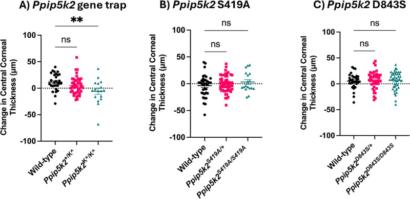
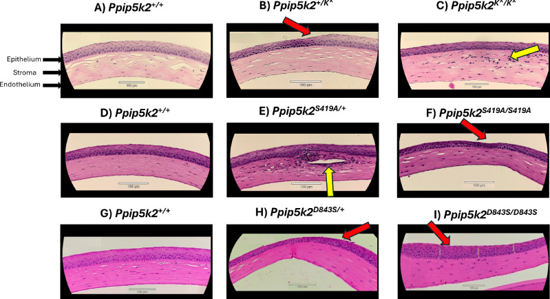
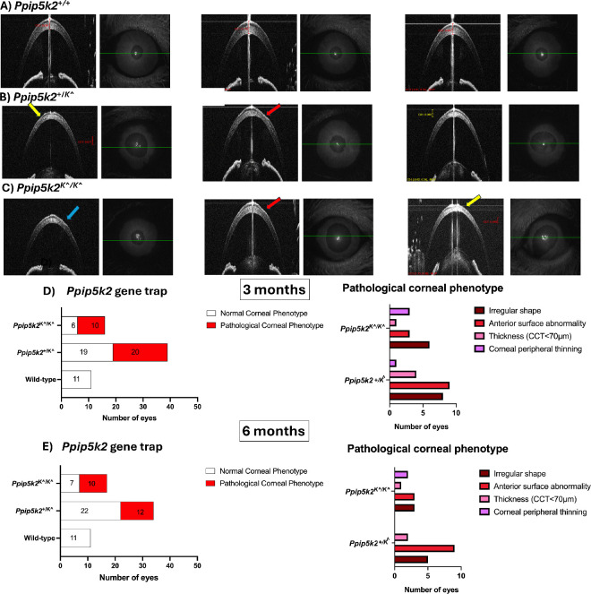
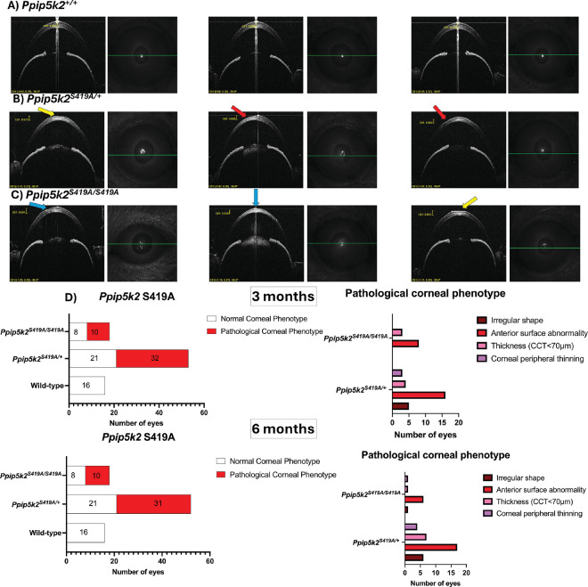
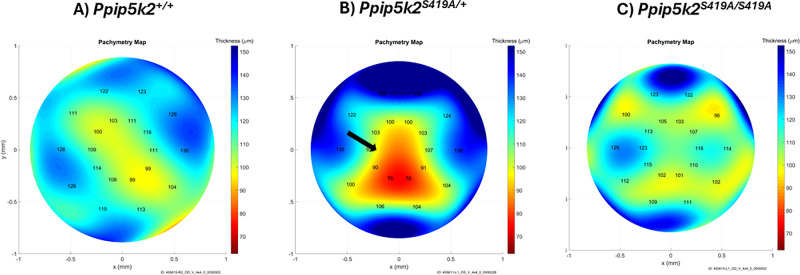
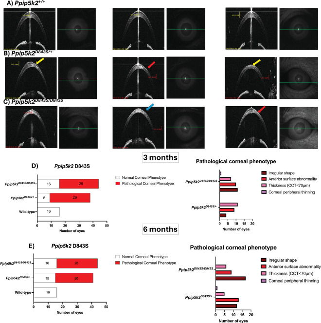
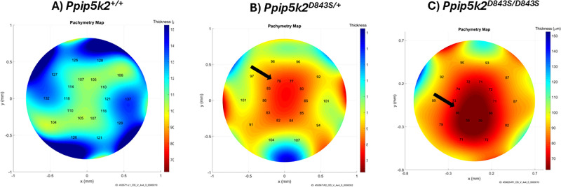
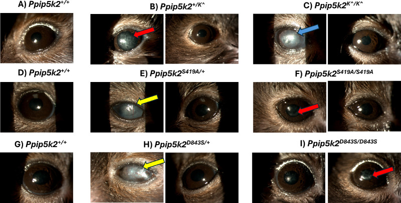
Central corneal thickness (CCT) was determined using spectral domain optical coherence tomography (SD-OCT) in Ppip5k2+/K^ (n = 41 eyes), Ppip5k2K^/K^ (n = 17 eyes) and 2 knock-in mice, Ppip5k2S419A/+ (n = 54 eyes) and Ppip5k2S419A/S419A (n = 18 eyes), and Ppip5k2D843S/+ (n = 42 eyes) and Ppip5k2D843S/D843S (n = 44 eyes) at 3 and 6 months. Pachymetry maps were generated using the Mouse Corneal Analysis Program (MCAP) to process OCT images. Slit lamp biomicroscopy was used to determine any corneal abnormalities, and, last, hematoxylin and eosin (H&E) staining using corneal sections from these animals was used to examine morphological changes.

RESULTS

CCT significantly decreased from 3 to 6 months in the Ppip5k2+/K^ and Ppip5k2K^/K^ mice compared to their littermate controls. OCT-based pachymetry maps revealed abnormally localized thinning in all three models compared to their wild-type (WT) controls. Slit lamp examinations revealed corneal abnormalities in the form of bullous keratopathy, stromal edema, stromal scarring, deep corneal neovascularization, and opacities in the heterozygous/homozygous mice of the three models in comparison with their controls. Corneal histological abnormalities, such as epithelial thickening and stromal layer damage, were observed in the heterozygous/homozygous mice of the three models in comparison with the WT controls.

CONCLUSIONS

We have identified phenotypic and histological changes in the corneas of three mouse lines that could be relevant in the development of animal models of KC.

摘要

目的

为了研究和治疗圆锥角膜(KC),建立一种小鼠模型是很有必要的。我们旨在确定3种Ppip5k2小鼠模型的角膜表型。

方法

使用光谱域光学相干断层扫描(SD-OCT)测定Ppip5k2+/K^(n = 41只眼)、Ppip5k2K^/K^(n = 17只眼)以及2种基因敲入小鼠Ppip5k2S419A/+(n = 54只眼)、Ppip5k2S419A/S419A(n = 18只眼)、Ppip5k2D843S/+(n = 42只眼)和Ppip5k2D843S/D843S(n = 44只眼)在3个月和6个月时的中央角膜厚度(CCT)。使用小鼠角膜分析程序(MCAP)生成测厚图来处理OCT图像。使用裂隙灯生物显微镜检查确定是否存在角膜异常,最后,对这些动物的角膜切片进行苏木精和伊红(H&E)染色以检查形态学变化。

结果

与同窝对照相比,Ppip5k2+/K^和Ppip5k2K^/K^小鼠的CCT在3个月至6个月时显著降低。基于OCT的测厚图显示,与野生型(WT)对照相比,所有三种模型均存在异常局限性变薄。裂隙灯检查显示,与对照相比,三种模型的杂合子/纯合子小鼠出现了大疱性角膜病变、基质水肿、基质瘢痕、角膜深层新生血管和混浊等角膜异常。与WT对照相比,在三种模型的杂合子/纯合子小鼠中观察到角膜组织学异常,如上皮增厚和基质层损伤。

结论

我们已经确定了三种小鼠品系角膜中的表型和组织学变化,这些变化可能与KC动物模型的开发相关。

https://cdn.ncbi.nlm.nih.gov/pmc/blobs/0dd0/11178121/911e9833013d/iovs-65-6-22-f009.jpg
https://cdn.ncbi.nlm.nih.gov/pmc/blobs/0dd0/11178121/0b5dae648957/iovs-65-6-22-f001.jpg
https://cdn.ncbi.nlm.nih.gov/pmc/blobs/0dd0/11178121/d455ab896ecc/iovs-65-6-22-f002.jpg
https://cdn.ncbi.nlm.nih.gov/pmc/blobs/0dd0/11178121/f926d22c7a9a/iovs-65-6-22-f003.jpg
https://cdn.ncbi.nlm.nih.gov/pmc/blobs/0dd0/11178121/a5541b555bd6/iovs-65-6-22-f004.jpg
https://cdn.ncbi.nlm.nih.gov/pmc/blobs/0dd0/11178121/74f09861117a/iovs-65-6-22-f005.jpg
https://cdn.ncbi.nlm.nih.gov/pmc/blobs/0dd0/11178121/856ea32e499b/iovs-65-6-22-f006.jpg
https://cdn.ncbi.nlm.nih.gov/pmc/blobs/0dd0/11178121/1b50ea2454ca/iovs-65-6-22-f007.jpg
https://cdn.ncbi.nlm.nih.gov/pmc/blobs/0dd0/11178121/d6f602a26c3d/iovs-65-6-22-f008.jpg
https://cdn.ncbi.nlm.nih.gov/pmc/blobs/0dd0/11178121/911e9833013d/iovs-65-6-22-f009.jpg

相似文献

1
Identification of Keratoconus-Related Phenotypes in Three Ppip5k2 Mouse Models.在三种Ppip5k2小鼠模型中鉴定圆锥角膜相关表型
Invest Ophthalmol Vis Sci. 2024 Jun 3;65(6):22. doi: 10.1167/iovs.65.6.22.
2
Comparison of corneal measurements in normal and keratoconus eyes using Anterior Segment Optical Coherence Tomography (AS-OCT) and Pentacam HR topographer.使用眼前节光学相干断层扫描(AS-OCT)和 Pentacam HR 地形图比较正常眼和圆锥角膜眼中的角膜测量值。
BMC Ophthalmol. 2023 May 3;23(1):194. doi: 10.1186/s12886-023-02946-w.
3
Fourier-domain optical coherence tomography imaging in keratoconus: a corneal structural classification.频域光学相干断层成像在圆锥角膜中的应用:角膜结构分类。
Ophthalmology. 2013 Dec;120(12):2403-2412. doi: 10.1016/j.ophtha.2013.05.027. Epub 2013 Aug 9.
4
Distinguishing Highly Asymmetric Keratoconus Eyes Using Combined Scheimpflug and Spectral-Domain OCT Analysis.使用联合 Scheimpflug 和频域 OCT 分析区分高度非对称圆锥角膜眼。
Ophthalmology. 2018 Dec;125(12):1862-1871. doi: 10.1016/j.ophtha.2018.06.020. Epub 2018 Jul 25.
5
A Coincident Thinning Index for Keratoconus Identification Using OCT Pachymetry and Epithelial Thickness Maps.使用 OCT 角膜地形图和上皮厚度图识别圆锥角膜的吻合性变薄指数。
J Refract Surg. 2020 Nov 1;36(11):757-765. doi: 10.3928/1081597X-20200925-01.
6
Corneal thickness in keratoconus: comparing optical, ultrasound, and optical coherence tomography pachymetry.圆锥角膜的角膜厚度:比较光学、超声和光学相干断层扫描测厚术。
Ophthalmology. 2013 Mar;120(3):457-463. doi: 10.1016/j.ophtha.2012.08.036. Epub 2012 Nov 21.
7
Differentiating Keratoconus and Corneal Warpage by Analyzing Focal Change Patterns in Corneal Topography, Pachymetry, and Epithelial Thickness Maps.通过分析角膜地形图、角膜厚度测量图和上皮厚度图中的焦点变化模式鉴别圆锥角膜和角膜弯曲度异常
Invest Ophthalmol Vis Sci. 2016 Jul 1;57(9):OCT544-9. doi: 10.1167/iovs.15-18938.
8
Corneal layer thickness in keratoconus using optical coherence tomography.角膜层厚度在圆锥角膜的光学相干断层扫描。
Clin Exp Optom. 2024 Jan;107(1):32-39. doi: 10.1080/08164622.2023.2201369. Epub 2023 Apr 30.
9
[Pachymetry with a video slit lamp].[使用视频裂隙灯进行眼压测量]
Ophthalmologe. 2021 Feb;118(2):162-168. doi: 10.1007/s00347-020-01108-3.
10
Corneal epithelial thickness mapping using Fourier-domain optical coherence tomography for detection of form fruste keratoconus.使用傅里叶域光学相干断层扫描进行角膜上皮厚度测绘以检测forme fruste圆锥角膜。
J Cataract Refract Surg. 2015 Apr;41(4):812-20. doi: 10.1016/j.jcrs.2014.06.043.

引用本文的文献

1
An Improved Monte Carlo Method for Quantitative Analysis of Transparency Degradation Caused by Corneal Edema.一种用于定量分析角膜水肿导致的透明度下降的改进蒙特卡罗方法。
Transl Vis Sci Technol. 2025 Apr 1;14(4):15. doi: 10.1167/tvst.14.4.15.

本文引用的文献

1
Animal Models for the Study of Keratoconus.用于研究圆锥角膜的动物模型。
Cells. 2023 Nov 22;12(23):2681. doi: 10.3390/cells12232681.
2
Inositol pyrophosphate dynamics reveals control of the yeast phosphate starvation program through 1,5-IP and the SPX domain of Pho81.焦磷酸肌醇动力学揭示了通过 1,5-IP 和 Pho81 的 SPX 结构域对酵母磷酸盐饥饿程序的控制。
Elife. 2023 Sep 20;12:RP87956. doi: 10.7554/eLife.87956.
3
Exosomes Released by Corneal Stromal Cells Show Molecular Alterations in Keratoconus Patients and Induce Different Cellular Behavior.
角膜基质细胞释放的外泌体在圆锥角膜患者中显示出分子改变并诱导不同的细胞行为。
Biomedicines. 2022 Sep 21;10(10):2348. doi: 10.3390/biomedicines10102348.
4
The Underlying Relationship between Keratoconus and Down Syndrome.圆锥角膜与唐氏综合征的潜在关系。
Int J Mol Sci. 2022 Sep 16;23(18):10796. doi: 10.3390/ijms231810796.
5
Keratoconus epidemiology: A review.圆锥角膜流行病学:综述
Saudi J Ophthalmol. 2022 Jul 11;36(1):3-6. doi: 10.4103/sjopt.sjopt_204_21. eCollection 2022 Jan-Mar.
6
Association of Novel Loci With Keratoconus Susceptibility in a Multitrait Genome-Wide Association Study of the UK Biobank Database and Canadian Longitudinal Study on Aging.在 UK Biobank 数据库和加拿大老龄化纵向研究的多性状全基因组关联研究中,与圆锥角膜易感性相关的新基因座的关联。
JAMA Ophthalmol. 2022 Jun 1;140(6):568-576. doi: 10.1001/jamaophthalmol.2022.0891.
7
Keratoconus: An updated review.圆锥角膜:更新综述。
Cont Lens Anterior Eye. 2022 Jun;45(3):101559. doi: 10.1016/j.clae.2021.101559. Epub 2022 Jan 4.
8
Anterior pituitary, sex hormones, and keratoconus: Beyond traditional targets.前垂体、性激素和圆锥角膜:超越传统靶点。
Prog Retin Eye Res. 2022 May;88:101016. doi: 10.1016/j.preteyeres.2021.101016. Epub 2021 Nov 2.
9
A multi-ethnic genome-wide association study implicates collagen matrix integrity and cell differentiation pathways in keratoconus.一项多民族全基因组关联研究表明圆锥角膜与胶原基质完整性及细胞分化途径有关。
Commun Biol. 2021 Mar 1;4(1):266. doi: 10.1038/s42003-021-01784-0.
10
Eye rubbing in the aetiology of keratoconus: a systematic review and meta-analysis.揉眼在圆锥角膜病因学中的作用:一项系统评价与荟萃分析。
Graefes Arch Clin Exp Ophthalmol. 2021 Aug;259(8):2057-2067. doi: 10.1007/s00417-021-05081-8. Epub 2021 Jan 23.