Buck Institute for Research on Aging, Novato, CA, USA.
USC Leonard Davis School of Gerontology, Los Angeles, CA, USA.
Geroscience. 2024 Oct;46(5):4185-4202. doi: 10.1007/s11357-024-01167-3. Epub 2024 Jun 13.
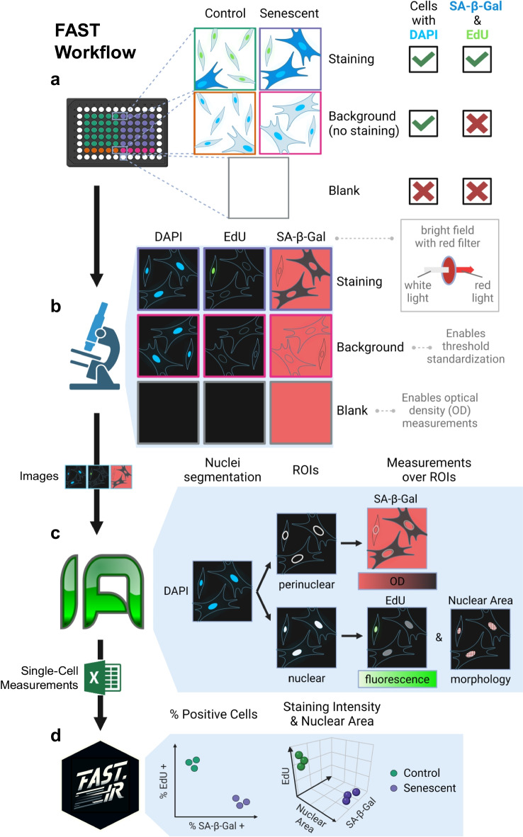
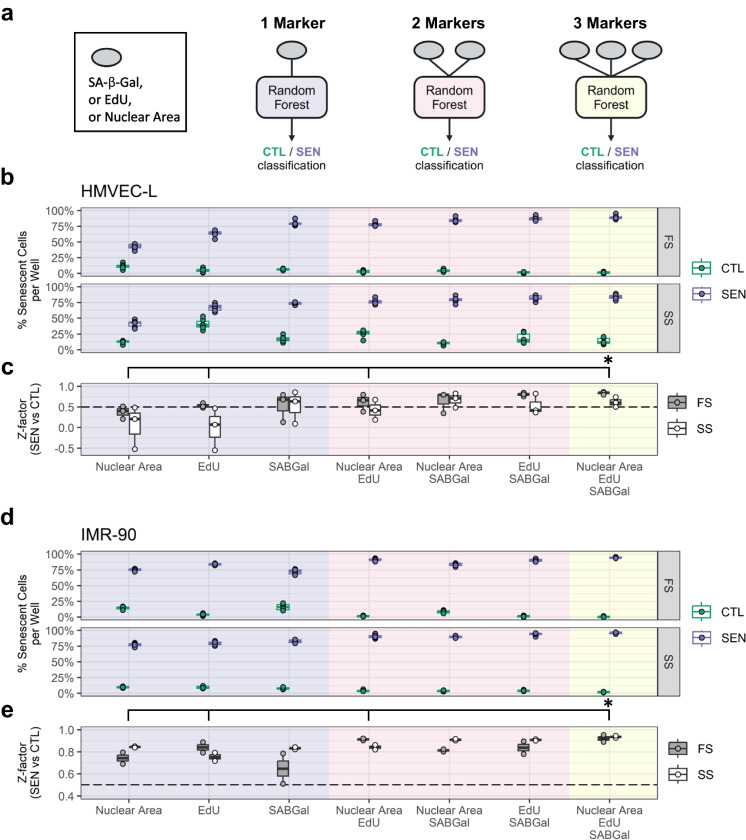
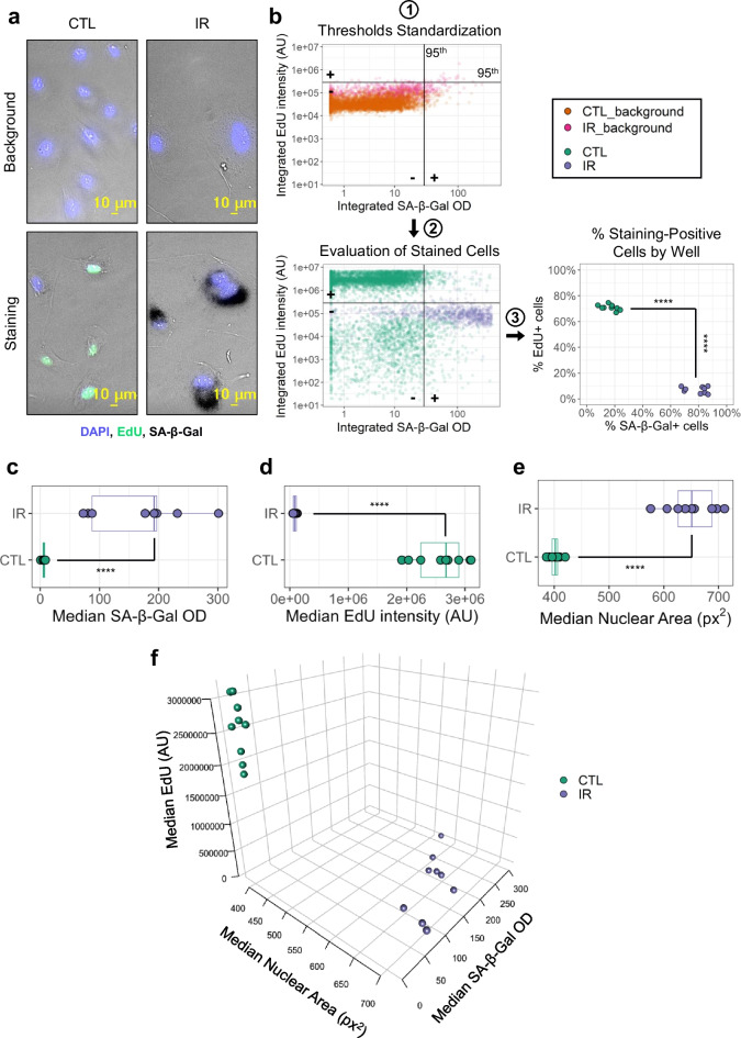
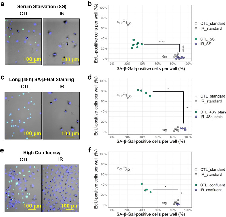
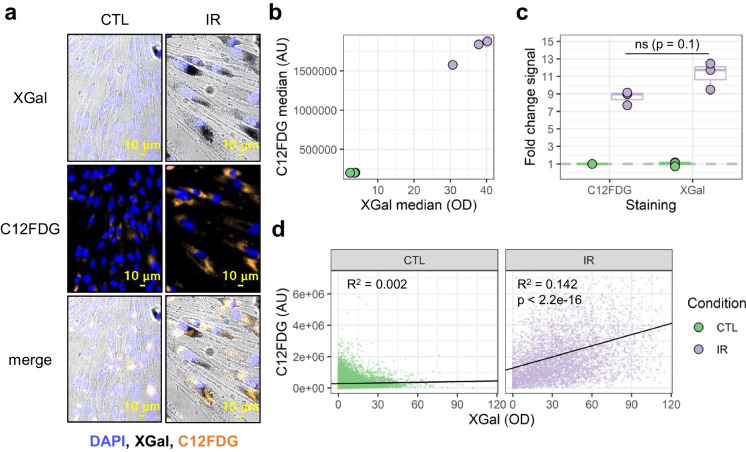
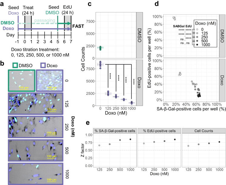
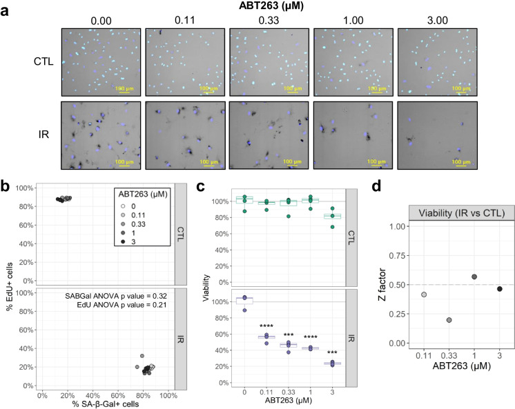
Cellular senescence is a major driver of aging and age-related diseases. Quantification of senescent cells remains challenging due to the lack of senescence-specific markers and generalist, unbiased methodology. Here, we describe the Fully-Automated Senescence Test (FAST), an image-based method for the high-throughput, single-cell assessment of senescence in cultured cells. FAST quantifies three of the most widely adopted senescence-associated markers for each cell imaged: senescence-associated β-galactosidase activity (SA-β-Gal) using X-Gal, proliferation arrest via lack of 5-ethynyl-2'-deoxyuridine (EdU) incorporation, and enlarged morphology via increased nuclear area. The presented workflow entails microplate image acquisition, image processing, data analysis, and graphing. Standardization was achieved by (i) quantifying colorimetric SA-β-Gal via optical density; (ii) implementing staining background controls; and (iii) automating image acquisition, image processing, and data analysis. In addition to the automated threshold-based scoring, a multivariate machine learning approach is provided. We show that FAST accurately quantifies senescence burden and is agnostic to cell type and microscope setup. Moreover, it effectively mitigates false-positive senescence marker staining, a common issue arising from culturing conditions. Using FAST, we compared X-Gal with fluorescent CFDG live-cell SA-β-Gal staining on the single-cell level. We observed only a modest correlation between the two, indicating that those stains are not trivially interchangeable. Finally, we provide proof of concept that our method is suitable for screening compounds that modify senescence burden. This method will be broadly useful to the aging field by enabling rapid, unbiased, and user-friendly quantification of senescence burden in culture, as well as facilitating large-scale experiments that were previously impractical.
细胞衰老(cellular senescence)是衰老和与年龄相关疾病的主要驱动因素。由于缺乏衰老特异性标志物和通用的、无偏的方法,衰老细胞的定量仍然具有挑战性。在这里,我们描述了一种全自动衰老测试(FAST),这是一种基于图像的方法,用于高通量、单细胞评估培养细胞中的衰老。FAST 量化了每个成像细胞中三种最广泛采用的衰老相关标志物:使用 X-Gal 的衰老相关β-半乳糖苷酶活性(SA-β-Gal)、缺乏 5-乙炔基-2'-脱氧尿苷(EdU)掺入的增殖停滞以及通过增加核面积的增大形态。呈现的工作流程包括微孔板图像采集、图像处理、数据分析和绘图。通过以下方法实现了标准化:(i)通过光密度量化比色法 SA-β-Gal;(ii)实施染色背景对照;(iii)自动化图像采集、图像处理和数据分析。除了自动化的基于阈值的评分外,还提供了一种多变量机器学习方法。我们表明 FAST 能够准确地定量衰老负担,并且与细胞类型和显微镜设置无关。此外,它有效地减轻了由于培养条件引起的假阳性衰老标志物染色的问题。使用 FAST,我们在单细胞水平上比较了 X-Gal 与荧光 CFDG 活细胞 SA-β-Gal 染色。我们观察到两者之间只有适度的相关性,这表明这些染色剂不能轻易互换。最后,我们提供了一个概念验证,证明我们的方法适用于筛选改变衰老负担的化合物。该方法将通过在培养物中快速、无偏和用户友好地定量衰老负担,以及促进以前不切实际的大规模实验,为衰老领域提供广泛的应用。