Department of Medical Ultrasonics, Guangxi Zhuang Autonomous Region, First Affiliated Hospital of Guangxi Medical University, Nanning, 530021, People's Republic of China.
Department of Medical Pathology, Guangxi Zhuang Autonomous Region, First Affiliated Hospital of Guangxi Medical University, Nanning, 530021, People's Republic of China.
Biomed Eng Online. 2024 Jun 18;23(1):56. doi: 10.1186/s12938-024-01259-3.
This study was designed to explore and validate the value of different machine learning models based on ultrasound image-omics features in the preoperative diagnosis of lymph node metastasis in pancreatic cancer (PC).
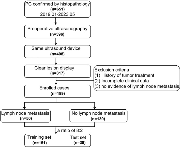
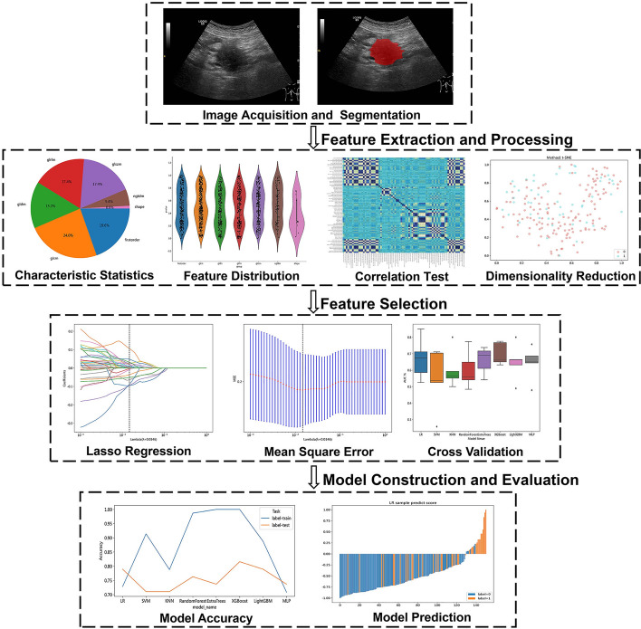
This research involved 189 individuals diagnosed with PC confirmed by surgical pathology (training cohort: n = 151; test cohort: n = 38), including 50 cases of lymph node metastasis. Image-omics features were extracted from ultrasound images. After dimensionality reduction and screening, eight machine learning algorithms, including logistic regression (LR), support vector machine (SVM), K-nearest neighbors (KNN), random forest (RF), extra trees (ET), extreme gradient boosting (XGBoost), light gradient boosting machine (LightGBM), and multilayer perceptron (MLP), were used to establish image-omics models to predict lymph node metastasis in PC. The best omics prediction model was selected through ROC curve analysis. Machine learning models were used to analyze clinical features and determine variables to establish a clinical model. A combined model was constructed by combining ultrasound image-omics and clinical features. Decision curve analysis (DCA) and a nomogram were used to evaluate the clinical application value of the model.
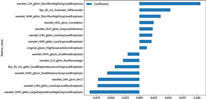
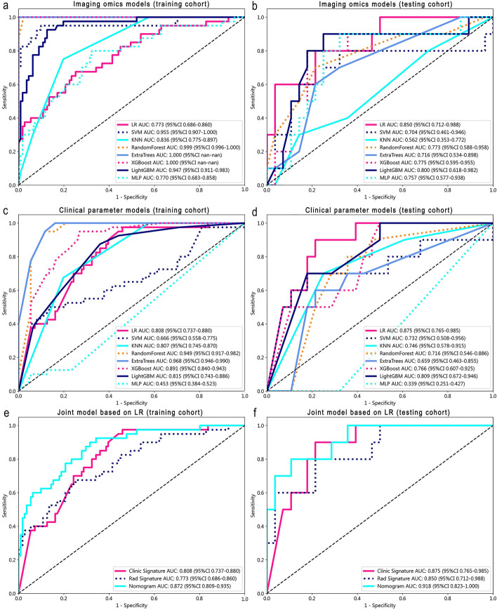
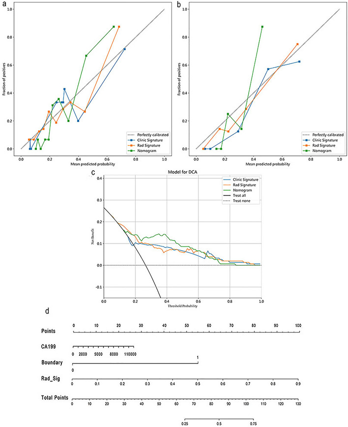
A total of 1561 image-omics features were extracted from ultrasound images. 15 valuable image-omics features were determined by regularization, dimension reduction, and algorithm selection. In the image-omics model, the LR model showed higher prediction efficiency and robustness, with an area under the ROC curve (AUC) of 0.773 in the training set and an AUC of 0.850 in the test set. The clinical model constructed by the boundary of lesions in ultrasound images and the clinical feature CA199 (AUC = 0.875). The combined model had the best prediction performance, with an AUC of 0.872 in the training set and 0.918 in the test set. The combined model showed better clinical benefit according to DCA, and the nomogram score provided clinical prediction solutions.
The combined model established with clinical features has good diagnostic ability and can be used to predict lymph node metastasis in patients with PC. It is expected to provide an effective noninvasive method for clinical decision-making, thereby improving the diagnosis and treatment of PC.
本研究旨在探索和验证基于超声影像组学特征的不同机器学习模型在术前诊断胰腺癌(PC)淋巴结转移中的价值。
本研究纳入了 189 名经手术病理证实为 PC 的患者(训练队列:n=151;测试队列:n=38),其中 50 例存在淋巴结转移。从超声图像中提取影像组学特征。经过降维和筛选,使用包括逻辑回归(LR)、支持向量机(SVM)、K 最近邻(KNN)、随机森林(RF)、极端梯度提升(XGBoost)、LightGBM、多层感知机(MLP)在内的 8 种机器学习算法建立预测 PC 淋巴结转移的影像组学模型。通过 ROC 曲线分析选择最佳的组学预测模型。使用机器学习模型分析临床特征并确定变量,建立临床模型。通过结合超声影像组学和临床特征构建联合模型。采用决策曲线分析(DCA)和列线图评估模型的临床应用价值。
从超声图像中提取了 1561 个影像组学特征。通过正则化、降维和算法选择,确定了 15 个有价值的影像组学特征。在影像组学模型中,LR 模型表现出更高的预测效率和稳健性,在训练集和测试集的 AUC 分别为 0.773 和 0.850。基于超声图像病灶边界和临床特征 CA199 构建的临床模型 AUC 为 0.875。联合模型的预测性能最佳,在训练集和测试集的 AUC 分别为 0.872 和 0.918。根据 DCA,联合模型显示出更好的临床获益,列线图评分提供了临床预测解决方案。
基于临床特征建立的联合模型具有良好的诊断能力,可用于预测 PC 患者的淋巴结转移。有望为临床决策提供一种有效的非侵入性方法,从而改善 PC 的诊断和治疗。