Koutarapu Srinivas, Ge Junyue, Dulewicz Maciej, Srikrishna Meera, Szadziewska Alicja, Wood Jack, Blennow Kaj, Zetterberg Henrik, Michno Wojciech, Ryan Natalie S, Lashley Tammaryn, Savas Jeffrey, Schöll Michael, Hanrieder Jörg
Department of Psychiatry and Neurochemistry, Sahlgrenska Academy, University of Gothenburg, Mölndal, Sweden.
Wallenberg Centre for Molecular and Translational Medicine, University of Gothenburg, Gothenburg, Sweden.
bioRxiv. 2024 Jun 3:2024.06.03.596890. doi: 10.1101/2024.06.03.596890.
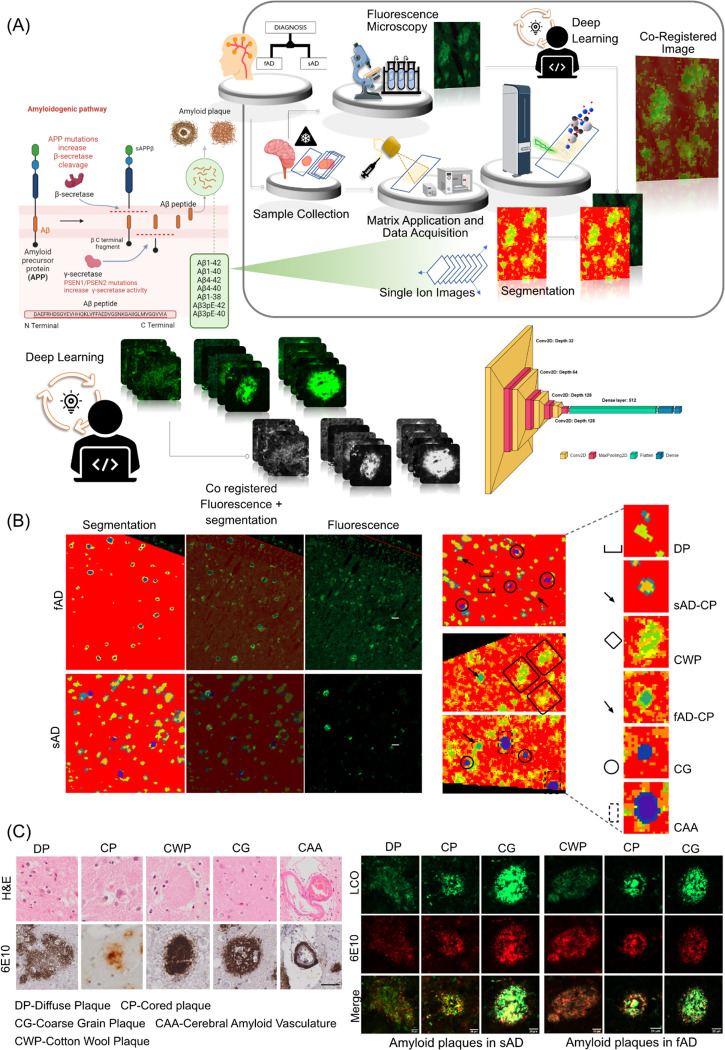
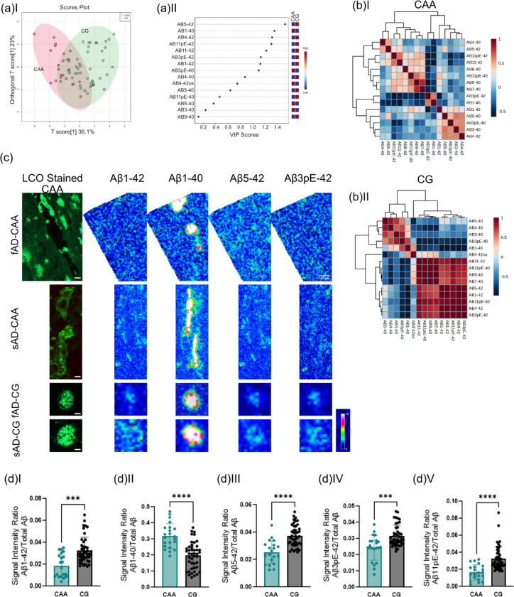
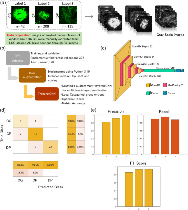
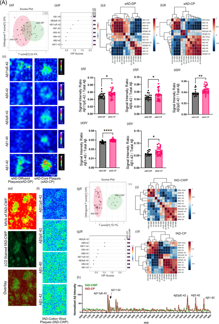
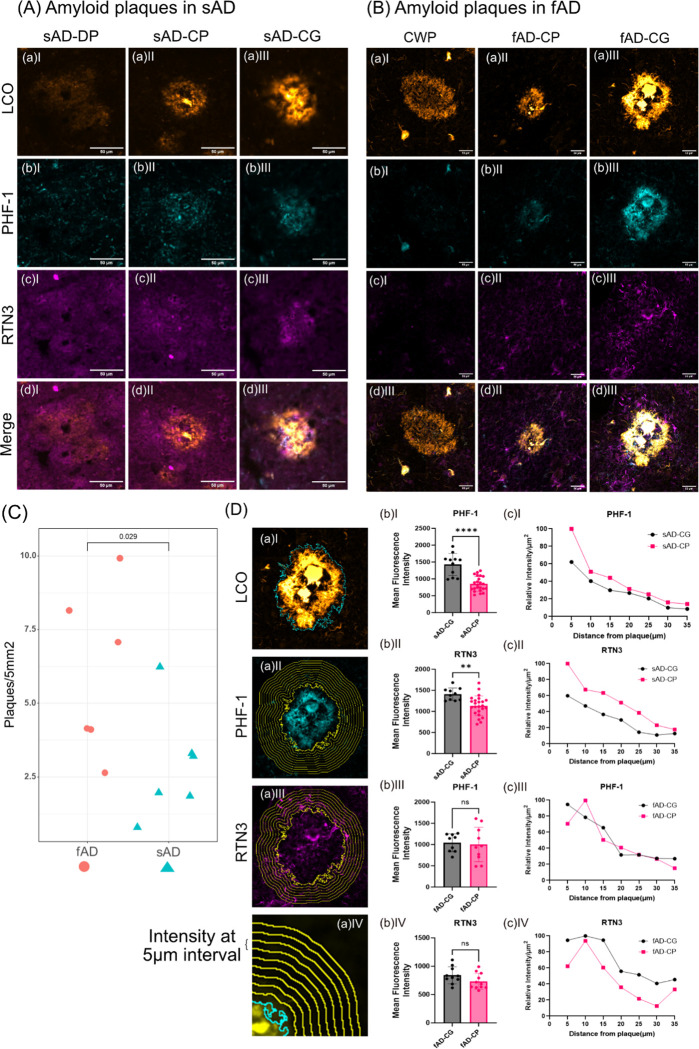
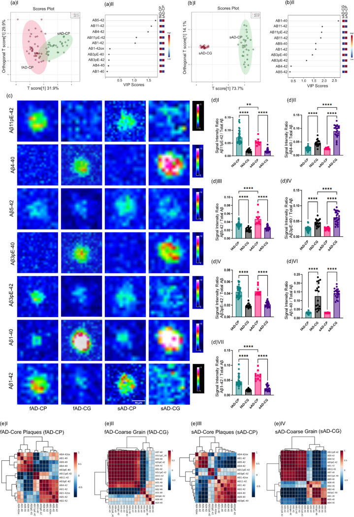
Amyloid plaque deposition is recognized as the primary pathological hallmark of Alzheimer's disease(AD) that precedes other pathological events and cognitive symptoms. Plaque pathology represents itself with an immense polymorphic variety comprising plaques with different stages of amyloid fibrillization ranging from diffuse to fibrillar, mature plaques. The association of polymorphic Aβ plaque pathology with AD pathogenesis, clinical symptoms and disease progression remains unclear. Advanced chemical imaging tools, such as functional amyloid microscopy combined with MALDI mass spectrometry imaging (MSI), are now enhanced by deep learning algorithms. This integration allows for precise delineation of polymorphic plaque structures and detailed identification of their associated Aβ compositions. We here set out to make use of these tools to interrogate heterogenic plaque types and their associated biochemical architecture. Our findings reveal distinct Aβ signatures that differentiate diffuse plaques from fibrilized ones, with the latter showing substantially higher levels of Aβx-40. Notably, within the fibrilized category, we identified a distinct subtype known as coarse-grain plaques. Both in sAD and fAD brain tissue, coarse grain plaques contained more Aβx-40 and less Aβx-42 compared with cored plaques. The coarse grain plaques in both sAD and fAD also showed higher levels of neuritic content including paired helical filaments (PHF-1)/phosphorylated phospho Tau-immunopositive neurites. Finally, the Aβ peptide content in coarse grain plaques resembled that of vascular Aβ deposits (CAA) though with relatively higher levels of Aβ1-42 and pyroglutamated Aβx-40 and Aβx-42 species in coarse grain plaques. This is the first of its kind study on spatial biochemical characterization of different plaque morphotypes demonstrating the potential of the correlative imaging techniques used that further increase the understanding of heterogeneous AD pathology. Linking the biochemical characteristics of amyloid plaque polymorphisms with various AD etiologies and toxicity mechanisms is crucial. Understanding the connection between plaque structure and disease pathogenesis can enhance our insights. This knowledge is particularly valuable for developing and advancing novel, amyloid-targeting therapeutics.
淀粉样斑块沉积被认为是阿尔茨海默病(AD)的主要病理标志,早于其他病理事件和认知症状。斑块病理学表现出巨大的多态性,包括从弥漫性到纤维状、成熟斑块等不同淀粉样纤维化阶段的斑块。多态性Aβ斑块病理学与AD发病机制、临床症状和疾病进展之间的关联仍不清楚。先进的化学成像工具,如功能淀粉样显微镜与基质辅助激光解吸电离质谱成像(MSI)相结合,现在通过深度学习算法得到了增强。这种整合允许精确描绘多态性斑块结构并详细识别其相关的Aβ组成。我们在此着手利用这些工具来研究异质性斑块类型及其相关的生化结构。我们的研究结果揭示了不同的Aβ特征,这些特征将弥漫性斑块与纤维化斑块区分开来,后者显示出显著更高水平的Aβx-40。值得注意的是,在纤维化类别中,我们鉴定出一种独特的亚型,称为粗颗粒斑块。在散发性AD(sAD)和家族性AD(fAD)脑组织中,与有核心斑块相比,粗颗粒斑块含有更多的Aβx-40和更少的Aβx-42。sAD和fAD中的粗颗粒斑块还显示出更高水平的神经突成分,包括双螺旋丝(PHF-1)/磷酸化磷酸化tau免疫阳性神经突。最后,粗颗粒斑块中的Aβ肽含量类似于血管Aβ沉积物(CAA),尽管粗颗粒斑块中Aβ1-42以及焦谷氨酸化Aβx-40和Aβx-42物种的水平相对较高。这是首次对不同斑块形态类型进行空间生化特征研究,证明了所使用的相关成像技术的潜力,进一步加深了对异质性AD病理学的理解。将淀粉样斑块多态性的生化特征与各种AD病因和毒性机制联系起来至关重要。理解斑块结构与疾病发病机制之间的联系可以增强我们的洞察力。这些知识对于开发和推进新型淀粉样蛋白靶向疗法特别有价值。