Eye Institute of Shandong First Medical University, Qingdao Eye Hospital of Shandong First Medical University, 5 Yan er dao Road, Qingdao, 266071, China.
State Key Laboratory Cultivation Base, Shandong Provincial Key Laboratory of Ophthalmology, Shandong First Medical University, Shandong, China.
BMC Ophthalmol. 2024 Jun 21;24(1):268. doi: 10.1186/s12886-024-03524-4.
Sleep deprivation (SD) is a common public health problem that contributes to various physiological disorders and increases the risk of ocular diseases. However, whether sleep loss can damage corneal endothelial function remains unclear. This study aimed to determine the effect and possible mechanism of SD on the corneal endothelium.
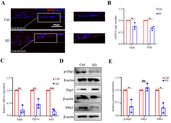
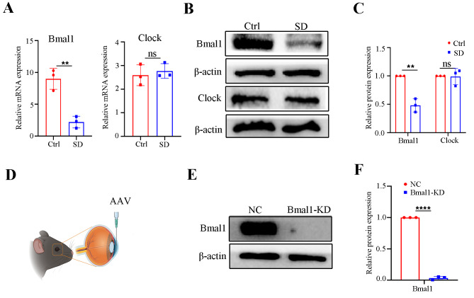
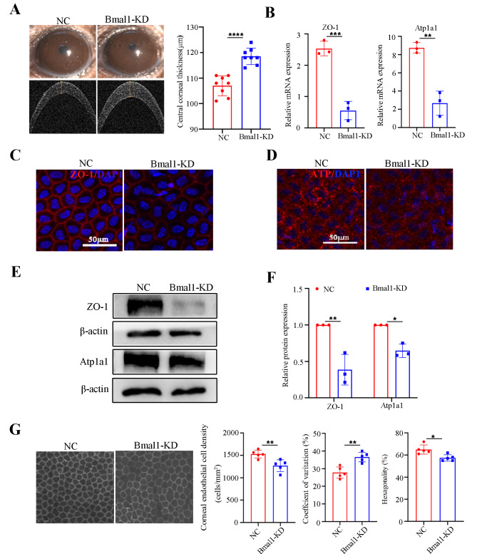
Male C57BL/6J mice were subjected to establish SD models. After 10 days, quantitative RT-PCR (qRT-PCR) and western blot or immunostaining for the expression levels of zonula occludens-1 (ZO-1), ATPase Na+/K + transporting subunit alpha 1 (Atp1a1), and core clock genes in the corneal endothelium were evaluated. Reactive oxygen species staining and mitochondrial abundance characterized the mitochondrial function. The regulatory role of Bmal1 was confirmed by specifically knocking down or overexpressing basic helix-loop-helix ARNT like 1 protein (Bmal1) in vivo. In vitro, a mitochondrial stress test was conducted on cultured human corneal endothelial cells upon Bmal1 knockdown.
SD damaged the barrier and pump functions of mouse corneal endothelium, accompanied by mitochondrial dysfunction. Interestingly, SD dramatically downregulated the core clock gene Bmal1 expression level. Bmal1 knockdown disrupted corneal endothelial function, while overexpression of Bmal1 ameliorated the dysfunction induced by SD. Mitochondrial bioenergetic deficiency mediated by Bmal1 was an underlying mechanism for SD induced corneal endothelial dysfunction.
The downregulation of Bmal1 expression caused by SD led to corneal endothelial dysfunction via impairing mitochondrial bioenergetics. Our findings offered insight into how SD impairs the physiological function of the corneal endothelium and expanded the understanding of sleep loss leading to ocular diseases.
睡眠剥夺(SD)是一种常见的公共卫生问题,可导致各种生理紊乱,并增加眼部疾病的风险。然而,睡眠不足是否会损害角膜内皮功能尚不清楚。本研究旨在确定 SD 对角膜内皮的影响及其可能的机制。
雄性 C57BL/6J 小鼠被用于建立 SD 模型。10 天后,评估角膜内皮中紧密连接蛋白-1(ZO-1)、ATP 酶 Na+/K+转运亚基α1(Atp1a1)和核心时钟基因的表达水平,采用定量 RT-PCR(qRT-PCR)和 Western blot 或免疫染色。通过活性氧染色和线粒体丰度来表征线粒体功能。通过体内特异性敲低或过表达基本螺旋-环-螺旋 ARNT 样 1 蛋白(Bmal1)来验证其调节作用。在体外,对培养的人角膜内皮细胞进行线粒体应激测试,以检测 Bmal1 敲低的影响。
SD 破坏了小鼠角膜内皮的屏障和泵功能,同时伴有线粒体功能障碍。有趣的是,SD 显著下调了核心时钟基因 Bmal1 的表达水平。Bmal1 敲低破坏了角膜内皮功能,而过表达 Bmal1 则改善了 SD 诱导的功能障碍。Bmal1 介导的线粒体生物能缺陷是 SD 诱导的角膜内皮功能障碍的潜在机制。
SD 导致 Bmal1 表达下调,通过损害线粒体生物能导致角膜内皮功能障碍。我们的研究结果为 SD 如何损害角膜内皮的生理功能提供了深入的了解,并扩展了对睡眠不足导致眼部疾病的认识。