Molecular Imaging Branch, National Cancer Institute, National Institutes of Health, 10 Center Dr., MSC 1182, Building 10, Room B3B85, Bethesda, MD, 20892, USA.
Department of Radiology, Singapore General Hospital, Singapore, Singapore.
Abdom Radiol (NY). 2024 Aug;49(8):2891-2901. doi: 10.1007/s00261-024-04468-5. Epub 2024 Jul 3.
To assess impact of image quality on prostate cancer extraprostatic extension (EPE) detection on MRI using a deep learning-based AI algorithm.
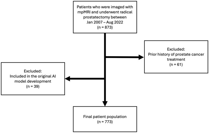
This retrospective, single institution study included patients who were imaged with mpMRI and subsequently underwent radical prostatectomy from June 2007 to August 2022. One genitourinary radiologist prospectively evaluated each patient using the NCI EPE grading system. Each T2WI was classified as low- or high-quality by a previously developed AI algorithm. Fisher's exact tests were performed to compare EPE detection metrics between low- and high-quality images. Univariable and multivariable analyses were conducted to assess the predictive value of image quality for pathological EPE.
A total of 773 consecutive patients (median age 61 [IQR 56-67] years) were evaluated. At radical prostatectomy, 23% (180/773) of patients had EPE at pathology, and 41% (131/318) of positive EPE calls on mpMRI were confirmed to have EPE. The AI algorithm classified 36% (280/773) of T2WIs as low-quality and 64% (493/773) as high-quality. For EPE grade ≥ 1, high-quality T2WI significantly improved specificity for EPE detection (72% [95% CI 67-76%] vs. 63% [95% CI 56-69%], P = 0.03), but did not significantly affect sensitivity (72% [95% CI 62-80%] vs. 75% [95% CI 63-85%]), positive predictive value (44% [95% CI 39-49%] vs. 38% [95% CI 32-43%]), or negative predictive value (89% [95% CI 86-92%] vs. 89% [95% CI 85-93%]). Sensitivity, specificity, PPV, and NPV for EPE grades ≥ 2 and ≥ 3 did not show significant differences attributable to imaging quality. For NCI EPE grade 1, high-quality images (OR 3.05, 95% CI 1.54-5.86; P < 0.001) demonstrated a stronger association with pathologic EPE than low-quality images (OR 1.76, 95% CI 0.63-4.24; P = 0.24).
Our study successfully employed a deep learning-based AI algorithm to classify image quality of prostate MRI and demonstrated that better quality T2WI was associated with more accurate prediction of EPE at final pathology.
使用基于深度学习的人工智能算法评估图像质量对前列腺癌前列腺外延伸(EPE)检测的影响。
本回顾性单中心研究纳入了 2007 年 6 月至 2022 年 8 月期间接受 mpMRI 检查并随后接受根治性前列腺切除术的患者。一名泌尿生殖放射科医生使用 NCI EPE 分级系统前瞻性地评估每位患者。使用先前开发的人工智能算法将每个 T2WI 分类为低质量或高质量。使用 Fisher 精确检验比较低质量和高质量图像之间的 EPE 检测指标。进行单变量和多变量分析,以评估图像质量对病理 EPE 的预测价值。
共评估了 773 例连续患者(中位年龄 61[IQR 56-67]岁)。在根治性前列腺切除术中,23%(180/773)的患者在病理上有 EPE,318 例 mpMRI 阳性 EPE 检查中有 41%(131/318)被证实有 EPE。人工智能算法将 36%(280/773)的 T2WI 分类为低质量,64%(493/773)为高质量。对于 EPE 分级≥1,高质量 T2WI 显著提高了 EPE 检测的特异性(72%[95%CI 67-76%]比 63%[95%CI 56-69%],P=0.03),但对敏感性(72%[95%CI 62-80%]比 75%[95%CI 63-85%])、阳性预测值(44%[95%CI 39-49%]比 38%[95%CI 32-43%])或阴性预测值(89%[95%CI 86-92%]比 89%[95%CI 85-93%])没有显著影响。EPE 分级≥2 和≥3 的敏感性、特异性、PPV 和 NPV 与图像质量无显著差异。对于 NCI EPE 分级 1,高质量图像(OR 3.05,95%CI 1.54-5.86;P<0.001)与低质量图像(OR 1.76,95%CI 0.63-4.24;P=0.24)相比,与病理 EPE 的关联更强。
本研究成功地使用基于深度学习的人工智能算法对前列腺 MRI 的图像质量进行分类,并表明质量更好的 T2WI 与更准确地预测最终病理 EPE 相关。