Department of Biomedical Engineering, Yale University, 10 Hillhouse Avenue, New Haven, CT, USA.
Systems Biology Institute, 850 West Campus Drive, West Haven, CT, USA.
Commun Biol. 2024 Jul 10;7(1):840. doi: 10.1038/s42003-024-06528-4.
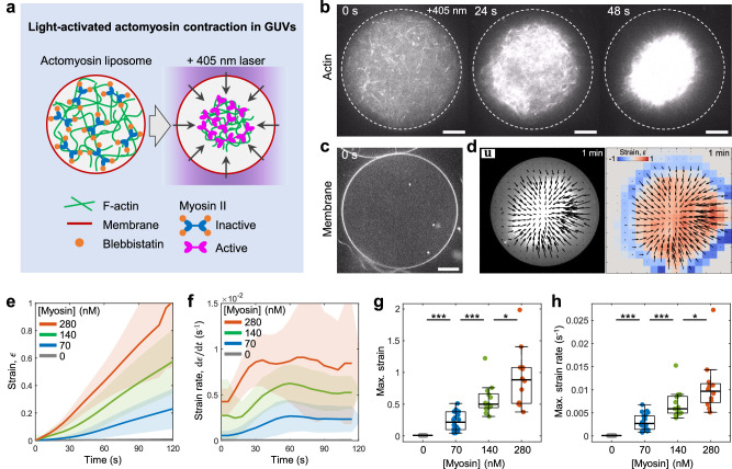
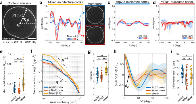
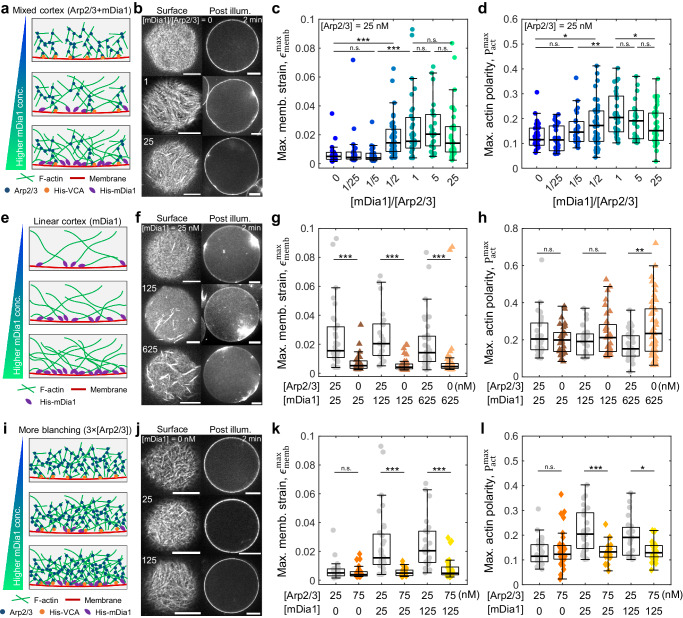
The architecture of the actin cortex determines the generation and transmission of stresses, during key events from cell division to migration. However, its impact on myosin-induced cell shape changes remains unclear. Here, we reconstitute a minimal model of the actomyosin cortex with branched or linear F-actin architecture within giant unilamellar vesicles (GUVs, liposomes). Upon light activation of myosin, neither the branched nor linear F-actin architecture alone induces significant liposome shape changes. The branched F-actin network forms an integrated, membrane-bound "no-slip boundary" -like cortex that attenuates actomyosin contractility. By contrast, the linear F-actin network forms an unintegrated "slip boundary" -like cortex, where actin asters form without inducing membrane deformations. Notably, liposomes undergo significant deformations at an optimized balance of branched and linear F-actin networks. Our findings highlight the pivotal roles of branched F-actin in force transmission and linear F-actin in force generation to yield membrane shape changes.
肌动蛋白皮层的结构决定了在细胞分裂到迁移等关键事件中力的产生和传递。然而,它对肌球蛋白诱导的细胞形状变化的影响尚不清楚。在这里,我们在巨大的单室脂质体(GUV,脂质体)中用分支或线性 F-肌动蛋白结构重建了一个最小的肌动球蛋白皮层模型。在肌球蛋白的光激活下,分支或线性 F-肌动蛋白结构本身都不会引起脂质体形状的显著变化。分支的 F-肌动蛋白网络形成一个整合的、膜结合的“无滑移边界”样皮层,从而减弱肌球蛋白收缩性。相比之下,线性 F-肌动蛋白网络形成一个未整合的“滑移边界”样皮层,其中肌动蛋白星状体形成而不会引起膜变形。值得注意的是,脂质体在分支和线性 F-肌动蛋白网络的优化平衡下会发生显著的变形。我们的发现强调了分支 F-肌动蛋白在力传递中的关键作用和线性 F-肌动蛋白在产生力以产生膜形状变化中的作用。