Institute of Diagnostic and Interventional Radiology and Neuroradiology, University Hospital Essen, Essen, Germany.
Institute for Artificial Intelligence in Medicine (IKIM), University Hospital Essen, Essen, Germany.
Crit Care. 2024 Jul 10;28(1):230. doi: 10.1186/s13054-024-05023-w.
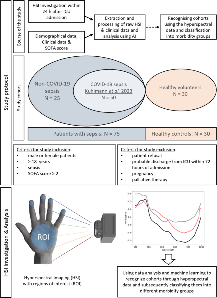
Impaired microcirculation is a cornerstone of sepsis development and leads to reduced tissue oxygenation, influenced by fluid and catecholamine administration during treatment. Hyperspectral imaging (HSI) is a non-invasive bedside technology for visualizing physicochemical tissue characteristics. Machine learning (ML) for skin HSI might offer an automated approach for bedside microcirculation assessment, providing an individualized tissue fingerprint of critically ill patients in intensive care. The study aimed to determine if machine learning could be utilized to automatically identify regions of interest (ROIs) in the hand, thereby distinguishing between healthy individuals and critically ill patients with sepsis using HSI.
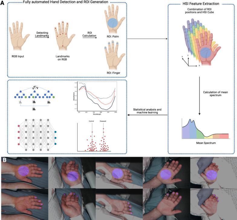
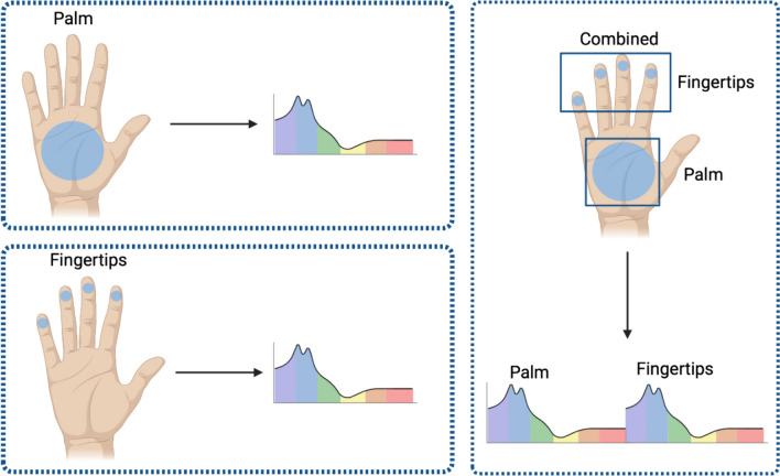
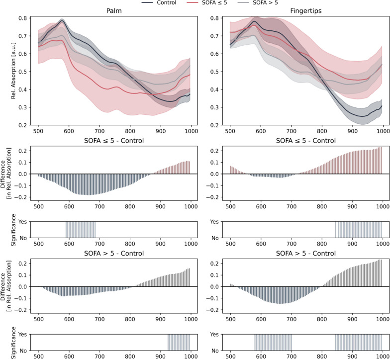
HSI raw data from 75 critically ill sepsis patients and from 30 healthy controls were recorded using TIVITA® Tissue System and analyzed using an automated ML approach. Additionally, patients were divided into two groups based on their SOFA scores for further subanalysis: less severely ill (SOFA ≤ 5) and severely ill (SOFA > 5). The analysis of the HSI raw data was fully-automated using MediaPipe for ROI detection (palm and fingertips) and feature extraction. HSI Features were statistically analyzed to highlight relevant wavelength combinations using Mann-Whitney-U test and Benjamini, Krieger, and Yekutieli (BKY) correction. In addition, Random Forest models were trained using bootstrapping, and feature importances were determined to gain insights regarding the wavelength importance for a model decision.
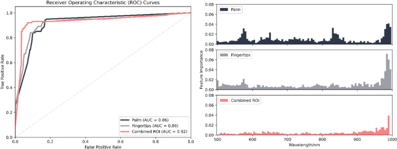
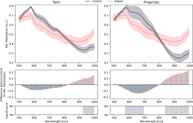
An automated pipeline for generating ROIs and HSI feature extraction was successfully established. HSI raw data analysis accurately distinguished healthy controls from sepsis patients. Wavelengths at the fingertips differed in the ranges of 575-695 nm and 840-1000 nm. For the palm, significant differences were observed in the range of 925-1000 nm. Feature importance plots indicated relevant information in the same wavelength ranges. Combining palm and fingertip analysis provided the highest reliability, with an AUC of 0.92 to distinguish between sepsis patients and healthy controls.
Based on this proof of concept, the integration of automated and standardized ROIs along with automated skin HSI analyzes, was able to differentiate between healthy individuals and patients with sepsis. This approach offers a reliable and objective assessment of skin microcirculation, facilitating the rapid identification of critically ill patients.
微循环受损是脓毒症发展的基石,导致组织氧合减少,这受到治疗过程中液体和儿茶酚胺给药的影响。高光谱成像(HSI)是一种非侵入性的床边技术,用于可视化理化组织特征。皮肤 HSI 的机器学习(ML)可能为床边微循环评估提供一种自动化方法,为重症监护中的危重病患者提供个性化的组织指纹。本研究旨在确定机器学习是否可用于自动识别手部的感兴趣区域(ROI),从而使用 HSI 区分健康个体和患有脓毒症的危重病患者。
使用 TIVITA® Tissue System 记录 75 名重症脓毒症患者和 30 名健康对照者的 HSI 原始数据,并使用自动 ML 方法进行分析。此外,根据 SOFA 评分将患者分为两组进行进一步的亚分析:病情较轻(SOFA≤5)和病情较重(SOFA>5)。使用 MediaPipe 对 HSI 原始数据进行全自动分析,以检测 ROI(手掌和指尖)并提取特征。使用 Mann-Whitney-U 检验和 Benjamini、Krieger 和 Yekutieli(BKY)校正对 HSI 特征进行统计学分析,以突出相关波长组合。此外,使用自举法训练随机森林模型,并确定特征重要性,以深入了解模型决策的波长重要性。
成功建立了生成 ROI 和 HSI 特征提取的自动化流水线。HSI 原始数据分析准确地区分了健康对照者和脓毒症患者。指尖处的波长在 575-695nm 和 840-1000nm 范围内存在差异。手掌处的波长在 925-1000nm 范围内存在显著差异。特征重要性图表明在相同的波长范围内存在相关信息。手掌和指尖分析相结合可提供最高的可靠性,区分脓毒症患者和健康对照者的 AUC 为 0.92。
基于这一概念验证,自动和标准化 ROI 的集成以及自动皮肤 HSI 分析能够区分健康个体和脓毒症患者。这种方法为皮肤微循环提供了可靠和客观的评估,有助于快速识别危重病患者。