• 文献检索
  • 文档翻译
  • 深度研究
  • 学术资讯
  • Suppr Zotero 插件Zotero 插件
  • 邀请有礼
  • 套餐&价格
  • 历史记录
应用&插件
Suppr Zotero 插件Zotero 插件浏览器插件Mac 客户端Windows 客户端微信小程序
定价
高级版会员购买积分包购买API积分包
服务
文献检索文档翻译深度研究API 文档MCP 服务
关于我们
关于 Suppr公司介绍联系我们用户协议隐私条款
关注我们

Suppr 超能文献

核心技术专利:CN118964589B侵权必究
粤ICP备2023148730 号-1Suppr @ 2026

文献检索

告别复杂PubMed语法,用中文像聊天一样搜索,搜遍4000万医学文献。AI智能推荐,让科研检索更轻松。

立即免费搜索

文件翻译

保留排版,准确专业,支持PDF/Word/PPT等文件格式,支持 12+语言互译。

免费翻译文档

深度研究

AI帮你快速写综述,25分钟生成高质量综述,智能提取关键信息,辅助科研写作。

立即免费体验

基于最小电极的高效广义脑电图瞌睡检测方法。

Efficient Generalized Electroencephalography-Based Drowsiness Detection Approach with Minimal Electrodes.

机构信息

Technology and Medical Imaging Laboratory, Faculty of Medicine Monastir, University of Monastir, Monastir 5019, Tunisia.

National Engineering School of Sousse, University of Sousse, BP 264 Erriyadh, Sousse 4023, Tunisia.

出版信息

Sensors (Basel). 2024 Jun 30;24(13):4256. doi: 10.3390/s24134256.

DOI:10.3390/s24134256
PMID:39001037
原文链接:https://pmc.ncbi.nlm.nih.gov/articles/PMC11244425/
Abstract

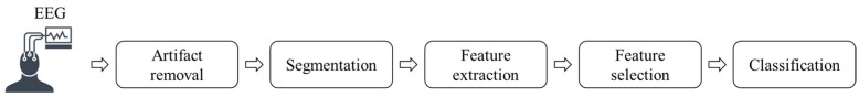
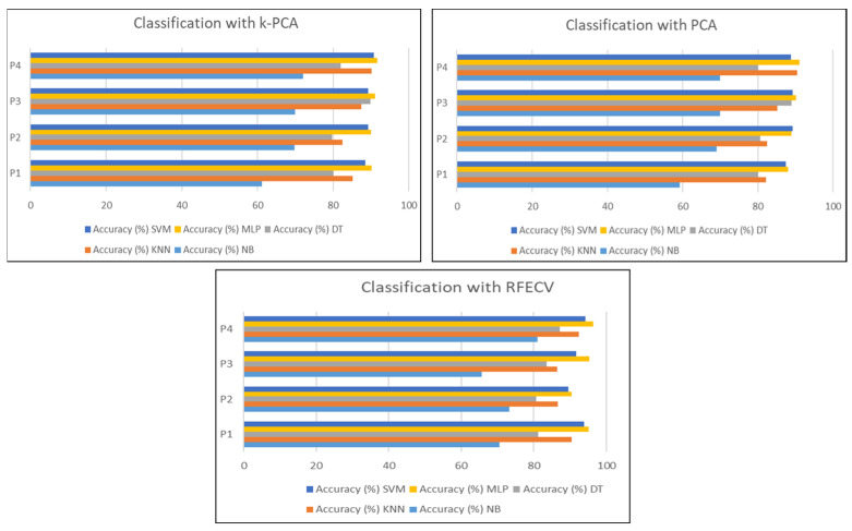
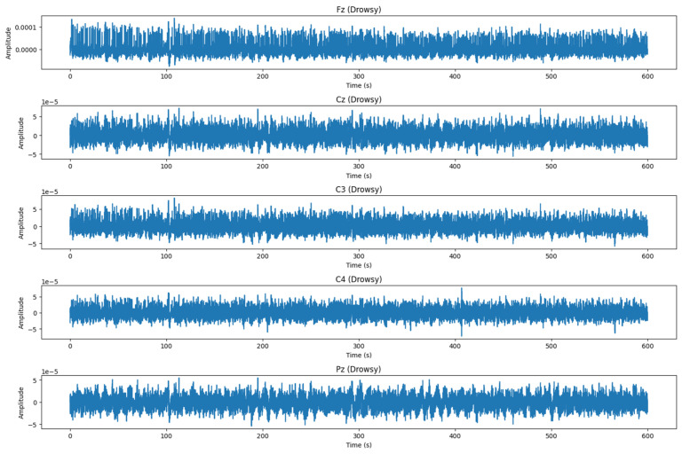
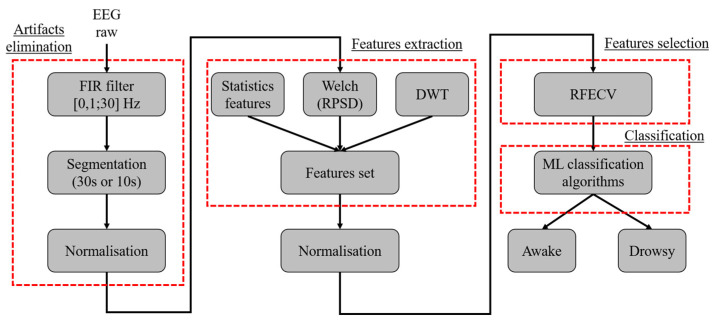
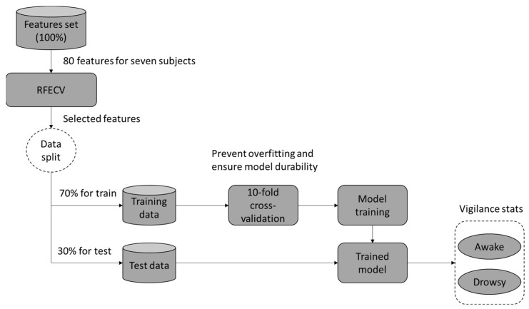
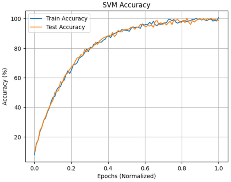
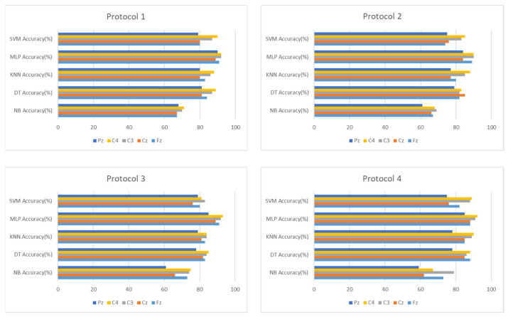
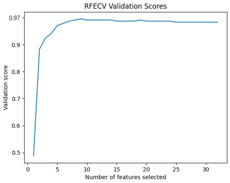
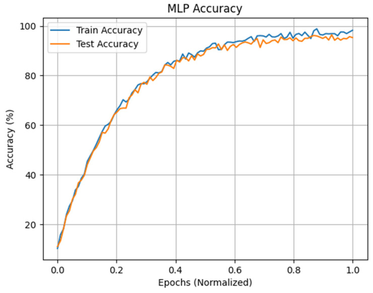
Drowsiness is a main factor for various costly defects, even fatal accidents in areas such as construction, transportation, industry and medicine, due to the lack of monitoring vigilance in the mentioned areas. The implementation of a drowsiness detection system can greatly help to reduce the defects and accident rates by alerting individuals when they enter a drowsy state. This research proposes an electroencephalography (EEG)-based approach for detecting drowsiness. EEG signals are passed through a preprocessing chain composed of artifact removal and segmentation to ensure accurate detection followed by different feature extraction methods to extract the different features related to drowsiness. This work explores the use of various machine learning algorithms such as Support Vector Machine (SVM), the K nearest neighbor (KNN), the Naive Bayes (NB), the Decision Tree (DT), and the Multilayer Perceptron (MLP) to analyze EEG signals sourced from the DROZY database, carefully labeled into two distinct states of alertness (awake and drowsy). Segmentation into 10 s intervals ensures precise detection, while a relevant feature selection layer enhances accuracy and generalizability. The proposed approach achieves high accuracy rates of 99.84% and 96.4% for intra (subject by subject) and inter (cross-subject) modes, respectively. SVM emerges as the most effective model for drowsiness detection in the intra mode, while MLP demonstrates superior accuracy in the inter mode. This research offers a promising avenue for implementing proactive drowsiness detection systems to enhance occupational safety across various industries.

摘要

困倦是导致建筑、交通、工业和医疗等领域各种代价高昂的缺陷甚至致命事故的主要因素,因为这些领域缺乏监测警觉性。实施困倦检测系统可以通过在个体进入困倦状态时发出警报,极大地有助于降低缺陷和事故率。本研究提出了一种基于脑电图(EEG)的困倦检测方法。EEG 信号通过预处理链(包括去除伪影和分段)进行处理,以确保准确检测,然后采用不同的特征提取方法提取与困倦相关的不同特征。这项工作探索了使用各种机器学习算法,如支持向量机(SVM)、K 最近邻(KNN)、朴素贝叶斯(NB)、决策树(DT)和多层感知机(MLP),来分析源自 DROZY 数据库的 EEG 信号,这些信号被仔细标记为两种不同的警觉状态(清醒和困倦)。将信号分段为 10 秒的间隔可确保精确检测,而相关的特征选择层可提高准确性和通用性。所提出的方法在内部(个体内)和外部(跨个体)模式下分别实现了 99.84%和 96.4%的高准确率。SVM 是内部模式下困倦检测最有效的模型,而 MLP 在外部模式下表现出更高的准确性。本研究为实施主动困倦检测系统提供了有前途的途径,可提高各个行业的职业安全水平。

https://cdn.ncbi.nlm.nih.gov/pmc/blobs/0281/11244425/842a3d2f3cef/sensors-24-04256-g017.jpg
https://cdn.ncbi.nlm.nih.gov/pmc/blobs/0281/11244425/866d11057d70/sensors-24-04256-g001.jpg
https://cdn.ncbi.nlm.nih.gov/pmc/blobs/0281/11244425/8f1acc6b6424/sensors-24-04256-g002.jpg
https://cdn.ncbi.nlm.nih.gov/pmc/blobs/0281/11244425/ee6de89554dc/sensors-24-04256-g003.jpg
https://cdn.ncbi.nlm.nih.gov/pmc/blobs/0281/11244425/8c7ef1c28ae8/sensors-24-04256-g004.jpg
https://cdn.ncbi.nlm.nih.gov/pmc/blobs/0281/11244425/305229e639ed/sensors-24-04256-g005.jpg
https://cdn.ncbi.nlm.nih.gov/pmc/blobs/0281/11244425/a3cca7dfafb9/sensors-24-04256-g006.jpg
https://cdn.ncbi.nlm.nih.gov/pmc/blobs/0281/11244425/fe38ae47e95d/sensors-24-04256-g007.jpg
https://cdn.ncbi.nlm.nih.gov/pmc/blobs/0281/11244425/036997dbf87b/sensors-24-04256-g008.jpg
https://cdn.ncbi.nlm.nih.gov/pmc/blobs/0281/11244425/c979d522af88/sensors-24-04256-g009.jpg
https://cdn.ncbi.nlm.nih.gov/pmc/blobs/0281/11244425/4204b46acfb6/sensors-24-04256-g010.jpg
https://cdn.ncbi.nlm.nih.gov/pmc/blobs/0281/11244425/b0dd015bc571/sensors-24-04256-g011.jpg
https://cdn.ncbi.nlm.nih.gov/pmc/blobs/0281/11244425/e2f9f83e477c/sensors-24-04256-g012.jpg
https://cdn.ncbi.nlm.nih.gov/pmc/blobs/0281/11244425/04cf7a9cce71/sensors-24-04256-g013.jpg
https://cdn.ncbi.nlm.nih.gov/pmc/blobs/0281/11244425/d0541477a5c6/sensors-24-04256-g014.jpg
https://cdn.ncbi.nlm.nih.gov/pmc/blobs/0281/11244425/ea0ecac28b58/sensors-24-04256-g015.jpg
https://cdn.ncbi.nlm.nih.gov/pmc/blobs/0281/11244425/0476b3455e00/sensors-24-04256-g016.jpg
https://cdn.ncbi.nlm.nih.gov/pmc/blobs/0281/11244425/842a3d2f3cef/sensors-24-04256-g017.jpg

相似文献

1
Efficient Generalized Electroencephalography-Based Drowsiness Detection Approach with Minimal Electrodes.基于最小电极的高效广义脑电图瞌睡检测方法。
Sensors (Basel). 2024 Jun 30;24(13):4256. doi: 10.3390/s24134256.
2
Drowsiness Detection Using Ocular Indices from EEG Signal.基于 EEG 信号的眼部指标的瞌睡检测。
Sensors (Basel). 2022 Jun 24;22(13):4764. doi: 10.3390/s22134764.
3
Drowsiness detection using portable wireless EEG.使用便携式无线 EEG 进行瞌睡检测。
Comput Methods Programs Biomed. 2022 Feb;214:106535. doi: 10.1016/j.cmpb.2021.106535. Epub 2021 Nov 16.
4
A Hybrid Approach to Detect Driver Drowsiness Utilizing Physiological Signals to Improve System Performance and Wearability.利用生理信号的混合方法检测驾驶员困倦,以提高系统性能和可穿戴性。
Sensors (Basel). 2017 Aug 31;17(9):1991. doi: 10.3390/s17091991.
5
Validation and interpretation of a multimodal drowsiness detection system using explainable machine learning.使用可解释机器学习验证和解释多模态瞌睡检测系统。
Comput Methods Programs Biomed. 2024 Jan;243:107925. doi: 10.1016/j.cmpb.2023.107925. Epub 2023 Nov 8.
6
Single-Channel Real-Time Drowsiness Detection Based on Electroencephalography.基于脑电图的单通道实时嗜睡检测
Annu Int Conf IEEE Eng Med Biol Soc. 2018 Jul;2018:98-101. doi: 10.1109/EMBC.2018.8512205.
7
Automatic classification methods for detecting drowsiness using wavelet packet transform extracted time-domain features from single-channel EEG signal.使用小波包变换从单通道脑电信号中提取时域特征的自动困倦检测分类方法。
J Neurosci Methods. 2021 Jan 1;347:108927. doi: 10.1016/j.jneumeth.2020.108927. Epub 2020 Sep 14.
8
An algorithm for automatic detection of drowsiness for use in wearable EEG systems.一种用于可穿戴脑电图系统的自动检测嗜睡的算法。
Annu Int Conf IEEE Eng Med Biol Soc. 2016 Aug;2016:3523-3526. doi: 10.1109/EMBC.2016.7591488.
9
Driving drowsiness detection using spectral signatures of EEG-based neurophysiology.基于脑电图神经生理学频谱特征的驾驶嗜睡检测
Front Physiol. 2023 Mar 30;14:1153268. doi: 10.3389/fphys.2023.1153268. eCollection 2023.
10
Portable Drowsiness Detection through Use of a Prefrontal Single-Channel Electroencephalogram.通过使用前额单通道脑电图进行便携性瞌睡检测。
Sensors (Basel). 2018 Dec 18;18(12):4477. doi: 10.3390/s18124477.

引用本文的文献

1
A Novel Hybrid Approach for Drowsiness Detection Using EEG Scalograms to Overcome Inter-Subject Variability.一种使用脑电图频谱图检测嗜睡的新型混合方法,以克服个体间差异。
Sensors (Basel). 2025 Sep 5;25(17):5530. doi: 10.3390/s25175530.

本文引用的文献

1
Ongoing EEG artifact correction using blind source separation.使用盲源分离进行持续 EEG 伪影校正。
Clin Neurophysiol. 2024 Feb;158:149-158. doi: 10.1016/j.clinph.2023.12.133. Epub 2024 Jan 4.
2
A systematic review of physiological signals based driver drowsiness detection systems.基于生理信号的驾驶员困倦检测系统的系统综述。
Cogn Neurodyn. 2023 Oct;17(5):1229-1259. doi: 10.1007/s11571-022-09898-9. Epub 2022 Oct 22.
3
Electroencephalography Signal Processing: A Comprehensive Review and Analysis of Methods and Techniques.
脑电图信号处理:方法和技术的综合回顾与分析。
Sensors (Basel). 2023 Jul 16;23(14):6434. doi: 10.3390/s23146434.
4
Real-Time Machine Learning-Based Driver Drowsiness Detection Using Visual Features.基于视觉特征的实时机器学习驾驶员困倦检测
J Imaging. 2023 Apr 29;9(5):91. doi: 10.3390/jimaging9050091.
5
Assessment of Vigilance Level during Work: Fitting a Hidden Markov Model to Heart Rate Variability.工作期间警觉水平的评估:将隐马尔可夫模型拟合于心率变异性
Brain Sci. 2023 Apr 7;13(4):638. doi: 10.3390/brainsci13040638.
6
Driving drowsiness detection using spectral signatures of EEG-based neurophysiology.基于脑电图神经生理学频谱特征的驾驶嗜睡检测
Front Physiol. 2023 Mar 30;14:1153268. doi: 10.3389/fphys.2023.1153268. eCollection 2023.
7
Designing an Embedded Feature Selection Algorithm for a Drowsiness Detector Model Based on Electroencephalogram Data.基于脑电数据的嵌入式特征选择算法设计用于瞌睡检测模型。
Sensors (Basel). 2023 Feb 7;23(4):1874. doi: 10.3390/s23041874.
8
System and Method for Driver Drowsiness Detection Using Behavioral and Sensor-Based Physiological Measures.基于行为和传感器的生理测量的驾驶员瞌睡检测系统和方法。
Sensors (Basel). 2023 Jan 23;23(3):1292. doi: 10.3390/s23031292.
9
Trends in EEG signal feature extraction applications.脑电图信号特征提取应用的趋势。
Front Artif Intell. 2023 Jan 25;5:1072801. doi: 10.3389/frai.2022.1072801. eCollection 2022.
10
Embedding decomposition for artifacts removal in EEG signals.用于去除脑电信号中伪迹的嵌入分解
J Neural Eng. 2022 Apr 22;19(2). doi: 10.1088/1741-2552/ac63eb.