Biodiscovery Institute, School of Medicine, University of Nottingham, Nottingham, NG7 2RD, UK.
Curr Oncol Rep. 2024 Oct;26(10):1213-1222. doi: 10.1007/s11912-024-01580-z. Epub 2024 Jul 16.
Isocitrate dehydrogenase wild-type glioblastoma is the most aggressive primary brain tumour in adults. Its infiltrative nature and heterogeneity confer a dismal prognosis, despite multimodal treatment. Precision medicine is increasingly advocated to improve survival rates in glioblastoma management; however, conventional neuroimaging techniques are insufficient in providing the detail required for accurate diagnosis of this complex condition.
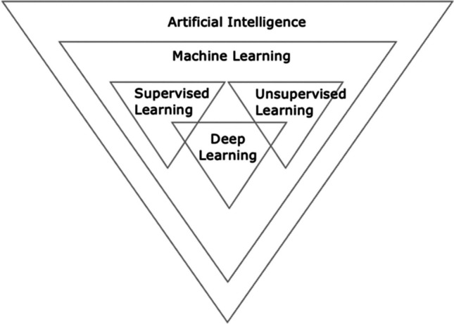
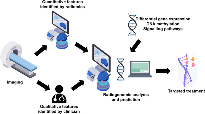
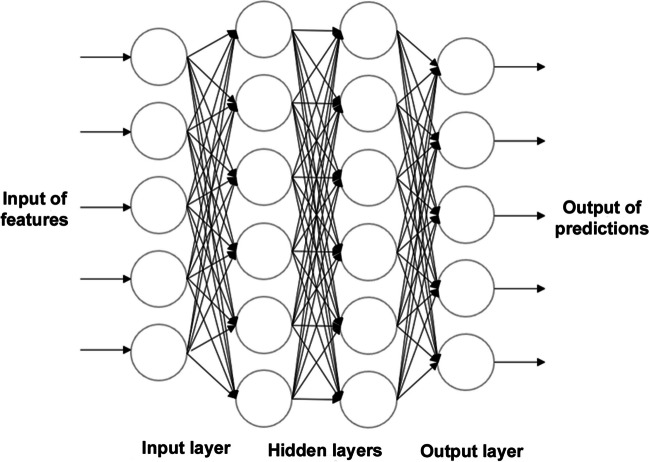
Advanced magnetic resonance imaging allows more comprehensive understanding of the tumour microenvironment. Combining diffusion and perfusion magnetic resonance imaging to create a multiparametric scan enhances diagnostic power and can overcome the unreliability of tumour characterisation by standard imaging. Recent progress in deep learning algorithms establishes their remarkable ability in image-recognition tasks. Integrating these with multiparametric scans could transform the diagnosis and monitoring of patients by ensuring that the entire tumour is captured. As a corollary, radiomics has emerged as a powerful approach to offer insights into diagnosis, prognosis, treatment, and tumour response through extraction of information from radiological scans, and transformation of these tumour characteristics into quantitative data. Radiogenomics, which links imaging features with genomic profiles, has exhibited its ability in characterising glioblastoma, and determining therapeutic response, with the potential to revolutionise management of glioblastoma. The integration of deep learning algorithms into radiogenomic models has established an automated, highly reproducible means to predict glioblastoma molecular signatures, further aiding prognosis and targeted therapy. However, challenges including lack of large cohorts, absence of standardised guidelines and the 'black-box' nature of deep learning algorithms, must first be overcome before this workflow can be applied in clinical practice.
异柠檬酸脱氢酶野生型脑胶质瘤是成人中最具侵袭性的原发性脑肿瘤。尽管采用了多模式治疗,但由于其浸润性和异质性,预后仍然很差。精准医学越来越被提倡用于提高脑胶质瘤治疗的生存率;然而,传统的神经影像学技术在提供准确诊断这种复杂疾病所需的细节方面是不够的。
高级磁共振成像可以更全面地了解肿瘤微环境。将扩散和灌注磁共振成像结合起来创建一个多参数扫描,可以提高诊断能力,并可以克服标准成像对肿瘤特征描述的不可靠性。深度学习算法的最新进展确立了它们在图像识别任务中的出色能力。将这些技术与多参数扫描相结合,可以通过确保整个肿瘤都被捕获,从而改变患者的诊断和监测方式。因此,放射组学已经成为一种强大的方法,可以通过从放射扫描中提取信息,并将这些肿瘤特征转化为定量数据,为诊断、预后、治疗和肿瘤反应提供深入的见解。放射基因组学将影像学特征与基因组图谱联系起来,已经显示出其在脑胶质瘤特征描述和治疗反应判断方面的能力,有可能彻底改变脑胶质瘤的治疗方法。将深度学习算法集成到放射基因组学模型中,建立了一种自动化、高度可重复的方法来预测脑胶质瘤的分子特征,进一步辅助预后和靶向治疗。然而,在这种工作流程能够应用于临床实践之前,必须首先克服缺乏大样本队列、缺乏标准化指南以及深度学习算法的“黑盒”性质等挑战。