• 文献检索
  • 文档翻译
  • 深度研究
  • 学术资讯
  • Suppr Zotero 插件Zotero 插件
  • 邀请有礼
  • 套餐&价格
  • 历史记录
应用&插件
Suppr Zotero 插件Zotero 插件浏览器插件Mac 客户端Windows 客户端微信小程序
定价
高级版会员购买积分包购买API积分包
服务
文献检索文档翻译深度研究API 文档MCP 服务
关于我们
关于 Suppr公司介绍联系我们用户协议隐私条款
关注我们

Suppr 超能文献

核心技术专利:CN118964589B侵权必究
粤ICP备2023148730 号-1Suppr @ 2026

文献检索

告别复杂PubMed语法,用中文像聊天一样搜索,搜遍4000万医学文献。AI智能推荐,让科研检索更轻松。

立即免费搜索

文件翻译

保留排版,准确专业,支持PDF/Word/PPT等文件格式,支持 12+语言互译。

免费翻译文档

深度研究

AI帮你快速写综述,25分钟生成高质量综述,智能提取关键信息,辅助科研写作。

立即免费体验

瘤牛(Bos Taurus indicus)头部:断层解剖与三维计算机断层扫描

Head of Zebu cattle (Bos Taurus indicus): sectional anatomy and 3D computed tomography.

作者信息

Nomir Ahmed G, El Sharaby Ashraf, Hanafy Basma G, Abumandour Mohamed M A

机构信息

Department of Anatomy and Embryology, Faculty of Veterinary Medicine, Damanhour University, Damanhour, 22511, Egypt.

Department of Anatomy and Embryology, Faculty of Veterinary Medicine, Alexandria University, Post Box: 22758, Alexandria, 21944, Egypt.

出版信息

BMC Vet Res. 2024 Jul 16;20(1):318. doi: 10.1186/s12917-024-04141-5.

DOI:10.1186/s12917-024-04141-5
PMID:39014413
原文链接:https://pmc.ncbi.nlm.nih.gov/articles/PMC11250968/
Abstract

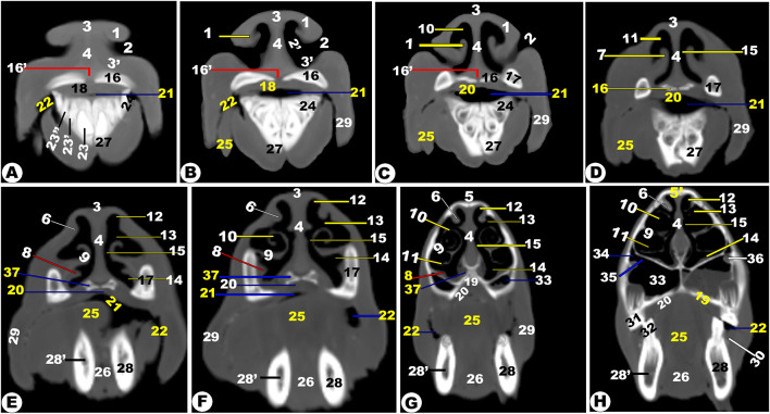
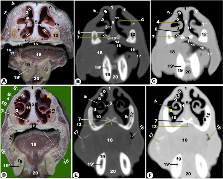
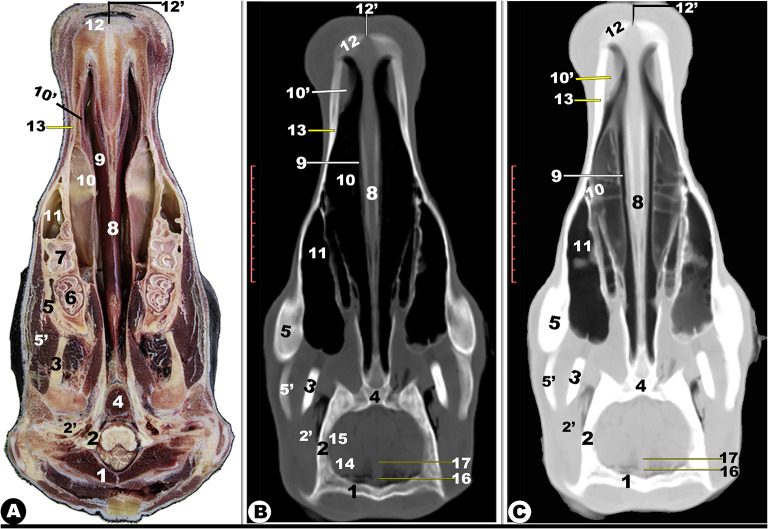
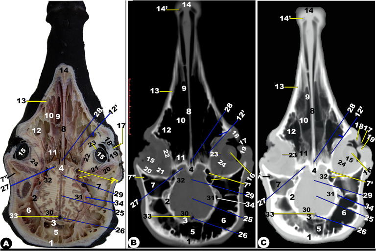
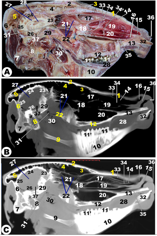
The research was designed to use computed tomography (CT) with 3D-CT reconstruction imaging techniques and the various anatomical sections-plana transversalia, frontalis, and dorsalia-to describe the anatomical architecture of the Zebu cattle head. Our study used nine mature heads. The CT bone window created detailed images of cranial bones, mandibles, teeth, and hyoid bones. All of the head cavities were evaluated, including the cranial, orbital, oral, auricular, and nasal cavities with their paranasal and conchal sinuses. The septum nasi, attached to the vomer and maxillary bones, did not reach the nasal cavity floor caudally at the level of the second premolar teeth, resulting in a single median channel from the choanae to the nasopharynx. The positions, boundaries, and connections of the paranasal sinuses were clearly identified. There were four nasal conchal sinuses (that were named the dorsal, middle, ethmoidal, and ventral) and five paranasal sinuses that were described as the following: sinus frontalis, maxillaris, palatinorum, and lacrimalis, as defined in the different anatomical sections and computed tomographic images. The complicated sinus frontalis caused the pneumatization of all bones that surrounded the cranial cavity, with the exception of the ethmoidal and body of basisphenoid bones. The sinus maxillaris was connected to the sinus lacrimalis and palatinorum through the maxillolacrimal and palatomaxillary openings, and to the middle nasal meatus through the nasomaxillary opening. Our findings provide a detailed anatomical knowledge for disease diagnosis to internal medicine veterinarians and surgeons by offering a comprehensive atlas of the Zebu cattle anatomy.

摘要

本研究旨在使用计算机断层扫描(CT)结合三维CT重建成像技术以及各种解剖切面——横断面、额面和背面——来描述瘤牛头的解剖结构。我们的研究使用了九个成熟的牛头。CT骨窗生成了颅骨、下颌骨、牙齿和舌骨的详细图像。对所有头部腔隙进行了评估,包括颅腔、眶腔、口腔、耳腔和鼻腔及其鼻窦和鼻甲窦。附着于犁骨和上颌骨的鼻中隔在第二前磨牙水平尾侧未到达鼻腔底部,导致从后鼻孔到鼻咽有一条单一的中通道。明确确定了鼻窦的位置、边界和连接。有四个鼻甲窦(分别命名为背侧、中间、筛窦和腹侧)和五个鼻窦,在不同的解剖切面和计算机断层图像中描述如下:额窦、上颌窦、腭窦和泪窦。复杂的额窦导致围绕颅腔的所有骨骼气化,但筛骨和蝶骨体除外。上颌窦通过上颌泪管开口和腭上颌开口与泪窦和腭窦相连,并通过鼻上颌开口与中鼻道相连。我们的研究结果通过提供瘤牛解剖结构的综合图谱,为内科兽医和外科医生的疾病诊断提供了详细的解剖学知识。

https://cdn.ncbi.nlm.nih.gov/pmc/blobs/d382/11250968/b30d9cac9fe6/12917_2024_4141_Fig15_HTML.jpg
https://cdn.ncbi.nlm.nih.gov/pmc/blobs/d382/11250968/eaba2010c88a/12917_2024_4141_Fig1_HTML.jpg
https://cdn.ncbi.nlm.nih.gov/pmc/blobs/d382/11250968/09affecb67f7/12917_2024_4141_Fig2_HTML.jpg
https://cdn.ncbi.nlm.nih.gov/pmc/blobs/d382/11250968/fabdaffc80b8/12917_2024_4141_Fig3_HTML.jpg
https://cdn.ncbi.nlm.nih.gov/pmc/blobs/d382/11250968/160cc24f5886/12917_2024_4141_Fig4_HTML.jpg
https://cdn.ncbi.nlm.nih.gov/pmc/blobs/d382/11250968/55b8fa26a9fc/12917_2024_4141_Fig5_HTML.jpg
https://cdn.ncbi.nlm.nih.gov/pmc/blobs/d382/11250968/e366024e7578/12917_2024_4141_Fig6_HTML.jpg
https://cdn.ncbi.nlm.nih.gov/pmc/blobs/d382/11250968/e366024e7578/12917_2024_4141_Fig7_HTML.jpg
https://cdn.ncbi.nlm.nih.gov/pmc/blobs/d382/11250968/5269885e1c87/12917_2024_4141_Fig8_HTML.jpg
https://cdn.ncbi.nlm.nih.gov/pmc/blobs/d382/11250968/8754433ec6b2/12917_2024_4141_Fig9_HTML.jpg
https://cdn.ncbi.nlm.nih.gov/pmc/blobs/d382/11250968/812ff9cf15e1/12917_2024_4141_Fig10_HTML.jpg
https://cdn.ncbi.nlm.nih.gov/pmc/blobs/d382/11250968/8e22c3b7bcda/12917_2024_4141_Fig11_HTML.jpg
https://cdn.ncbi.nlm.nih.gov/pmc/blobs/d382/11250968/aaa7f37d694b/12917_2024_4141_Fig12_HTML.jpg
https://cdn.ncbi.nlm.nih.gov/pmc/blobs/d382/11250968/3ce532b349d6/12917_2024_4141_Fig13_HTML.jpg
https://cdn.ncbi.nlm.nih.gov/pmc/blobs/d382/11250968/6c43ca040440/12917_2024_4141_Fig14_HTML.jpg
https://cdn.ncbi.nlm.nih.gov/pmc/blobs/d382/11250968/b30d9cac9fe6/12917_2024_4141_Fig15_HTML.jpg

相似文献

1
Head of Zebu cattle (Bos Taurus indicus): sectional anatomy and 3D computed tomography.瘤牛(Bos Taurus indicus)头部:断层解剖与三维计算机断层扫描
BMC Vet Res. 2024 Jul 16;20(1):318. doi: 10.1186/s12917-024-04141-5.
2
CT and cross-sectional anatomy of the paranasal sinuses in the Holstein cow.荷斯坦奶牛鼻窦的 CT 和横断解剖学。
Vet Radiol Ultrasound. 2023 Mar;64(2):211-223. doi: 10.1111/vru.13166. Epub 2022 Oct 20.
3
Anatomical description of the head in Ossimi Sheep: Sectional anatomy and Computed Tomographic approach.Ossimi 绵羊头部解剖结构描述:剖面解剖学和计算机断层扫描方法。
Morphologie. 2021 Feb;105(348):29-44. doi: 10.1016/j.morpho.2020.06.008. Epub 2020 Jul 6.
4
Anatomy of the head in the Saanen goat: a computed tomographic and cross-sectional approach.萨能山羊头部的解剖结构:计算机断层扫描和横断面研究方法。
Anat Sci Int. 2020 Jun;95(3):408-419. doi: 10.1007/s12565-020-00536-0. Epub 2020 Mar 11.
5
Computed tomography and sectional anatomy of the head cavities in donkey (Equus asinus).驴(马属驴)头腔的计算机断层扫描与断层解剖学
Anat Sci Int. 2014 Jun;89(3):140-50. doi: 10.1007/s12565-013-0209-7. Epub 2013 Oct 16.
6
Computed tomography and gross anatomical studies on the head of one-humped camel (Camelus dromedarius).单峰骆驼(骆驼属单峰驼)头部的计算机断层扫描和大体解剖学研究。
Anat Rec (Hoboken). 2014 Apr;297(4):630-42. doi: 10.1002/ar.22865. Epub 2014 Jan 13.
7
Volumetric measurements of paranasal sinuses and examination of sinonasal communication in healthy Shetland ponies: anatomical and morphometric characteristics using computed tomography.健康设得兰矮种马的副鼻窦容积测量和鼻旁窦沟通检查:使用计算机断层扫描的解剖学和形态测量特征。
BMC Vet Res. 2021 Jan 21;17(1):41. doi: 10.1186/s12917-021-02748-6.
8
The sinonasal communication in the horse: examinations using computerized three-dimensional reformatted renderings of computed-tomography datasets.马的鼻窦连通性:使用计算机断层扫描数据集的计算机三维重建图像进行的检查
BMC Vet Res. 2014 Mar 19;10:72. doi: 10.1186/1746-6148-10-72.
9
Using semi-automated segmentation of computed tomography datasets for three-dimensional visualization and volume measurements of equine paranasal sinuses.使用计算机断层扫描数据集的半自动分割技术对马鼻窦进行三维可视化和体积测量。
Vet Radiol Ultrasound. 2013 Nov-Dec;54(6):582-90. doi: 10.1111/vru.12080. Epub 2013 Jul 26.
10
Macroscopic anatomy, radiography and computed tomography of normal paranasal sinuses of the adult one-humped dromedary (Camelus dromedarius).成年单峰驼(Camelus dromedarius)正常鼻窦的大体解剖、X 线摄影和计算机断层扫描。
Vet Med Sci. 2021 Sep;7(5):1460-1468. doi: 10.1002/vms3.515. Epub 2021 May 1.

本文引用的文献

1
Anatomical studies on the PES region of Zebu cattle (Bos Taurus indicus) with special references to 3D computed tomography imaging technique.泽布牛(Bos Taurus indicus)PES 区域的解剖学研究,特别参考了 3D 计算机断层扫描成像技术。
BMC Vet Res. 2024 Mar 8;20(1):87. doi: 10.1186/s12917-024-03940-0.
2
Anatomy and computed tomography of the nasal cavity, nasal conchae, and paranasal sinuses of the endangered Patagonian huemul deer (Hippocamelus bisulcus).濒危巴塔哥尼亚南美泽鹿(Hippocamelus bisulcus)鼻腔、鼻甲及鼻窦的解剖结构与计算机断层扫描
Anat Rec (Hoboken). 2024 Jan;307(1):141-154. doi: 10.1002/ar.25230. Epub 2023 Apr 21.
3
CT and cross-sectional anatomy of the paranasal sinuses in the Holstein cow.
荷斯坦奶牛鼻窦的 CT 和横断解剖学。
Vet Radiol Ultrasound. 2023 Mar;64(2):211-223. doi: 10.1111/vru.13166. Epub 2022 Oct 20.
4
CT anatomy of the head in the Ile de France sheep.法兰西岛羊的头部 CT 解剖结构
Vet Med Sci. 2022 Jul;8(4):1694-1708. doi: 10.1002/vms3.834. Epub 2022 May 31.
5
Description of normal head structures of the one-humped camel () by magnetic resonance imaging, computed tomography, and cross-sectional anatomy.通过磁共振成像、计算机断层扫描和横断面解剖对单峰骆驼()正常头部结构的描述。
Vet World. 2020 Aug;13(8):1581-1587. doi: 10.14202/vetworld.2020.1581-1587. Epub 2020 Aug 14.
6
Anatomical description of the head in Ossimi Sheep: Sectional anatomy and Computed Tomographic approach.Ossimi 绵羊头部解剖结构描述:剖面解剖学和计算机断层扫描方法。
Morphologie. 2021 Feb;105(348):29-44. doi: 10.1016/j.morpho.2020.06.008. Epub 2020 Jul 6.
7
Anatomy of the head in the Saanen goat: a computed tomographic and cross-sectional approach.萨能山羊头部的解剖结构:计算机断层扫描和横断面研究方法。
Anat Sci Int. 2020 Jun;95(3):408-419. doi: 10.1007/s12565-020-00536-0. Epub 2020 Mar 11.
8
Surgical anatomy of the nasal and paranasal sinuses in Egyptian native sheep (Ovis aries) using computed tomography and cross sectioning.利用计算机断层扫描和横断面解剖对埃及本土绵羊(Ovis aries)鼻腔和鼻窦的外科解剖学研究
Anat Histol Embryol. 2019 Jul;48(4):279-289. doi: 10.1111/ahe.12436. Epub 2019 Apr 7.
9
Reconstruction and morphometric analysis of the nasal airway of the white-tailed deer (Odocoileus virginianus) and implications regarding respiratory and olfactory airflow.白尾鹿(弗吉尼亚鹿)鼻气道的重建与形态计量分析及其对呼吸气流和嗅觉气流的影响
Anat Rec (Hoboken). 2014 Nov;297(11):2138-47. doi: 10.1002/ar.23037.
10
Computed tomography and gross anatomical studies on the head of one-humped camel (Camelus dromedarius).单峰骆驼(骆驼属单峰驼)头部的计算机断层扫描和大体解剖学研究。
Anat Rec (Hoboken). 2014 Apr;297(4):630-42. doi: 10.1002/ar.22865. Epub 2014 Jan 13.