• 文献检索
  • 文档翻译
  • 深度研究
  • 学术资讯
  • Suppr Zotero 插件Zotero 插件
  • 邀请有礼
  • 套餐&价格
  • 历史记录
应用&插件
Suppr Zotero 插件Zotero 插件浏览器插件Mac 客户端Windows 客户端微信小程序
定价
高级版会员购买积分包购买API积分包
服务
文献检索文档翻译深度研究API 文档MCP 服务
关于我们
关于 Suppr公司介绍联系我们用户协议隐私条款
关注我们

Suppr 超能文献

核心技术专利:CN118964589B侵权必究
粤ICP备2023148730 号-1Suppr @ 2026

文献检索

告别复杂PubMed语法,用中文像聊天一样搜索,搜遍4000万医学文献。AI智能推荐,让科研检索更轻松。

立即免费搜索

文件翻译

保留排版,准确专业,支持PDF/Word/PPT等文件格式,支持 12+语言互译。

免费翻译文档

深度研究

AI帮你快速写综述,25分钟生成高质量综述,智能提取关键信息,辅助科研写作。

立即免费体验

法兰西岛羊的头部 CT 解剖结构

CT anatomy of the head in the Ile de France sheep.

机构信息

Department of Surgery and Radiology, Faculty of Veterinary Medicine, University of Tehran, Tehran, Iran.

Department of Basic Sciences, Faculty of Veterinary Medicine, University of Tehran, Tehran, Iran.

出版信息

Vet Med Sci. 2022 Jul;8(4):1694-1708. doi: 10.1002/vms3.834. Epub 2022 May 31.

DOI:10.1002/vms3.834
PMID:35639620
原文链接:https://pmc.ncbi.nlm.nih.gov/articles/PMC9297795/
Abstract

BACKGROUND

CT scan images provide accurate anatomical data from different areas of the body that can be used to diagnose diseases.

OBJECTIVES

The present work aimed to describe the normal anatomical structures of the Ile de France sheep head and its morphometric and volumetric properties using computed tomography (CT) and stereological methods.

METHODS

Five adult Ile de France sheep heads, which were of mature age (above 10 months), were included in this study. The different cavities of the head, including the nasal cavity, paranasal sinuses, oral cavity, orbital cavity and vestibulocochlear system, were evaluated using CT scans, cross, sagittal and coronal sections.

RESULTS

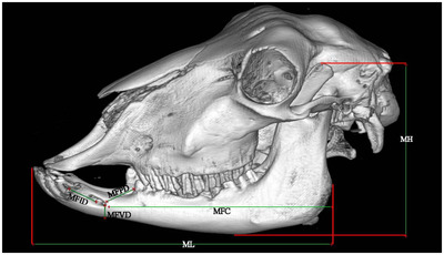
The mean length, height and width of the skull were 25.3 ± 1.02, 9.8 ± 0.93 and 12.3 ± 0.91 cm, respectively. The results showed that the nasal cavity is divided into three regions. Vestibular, respiratory and olfactory regions. The paranasal sinuses are composed of maxillary, frontal, palatine, sphenoid, lacrimal and ethmoidal that were identified and named in the CT scan images and their corresponding anatomical cross-sections. The total volume of the head, nasal cavity and oral cavity was estimated to be 2998 ± 202.00, 303 ± 31.33 and 229.3 ± 10.61 cm , respectively.

CONCLUSIONS

The frontal sinus in the Ile de France sheep was limited to the frontal bone without extending into the parietal, temporal, or occipital bones, similar to Saanen goat. This study provided a comprehensive atlas of Ile de France sheep anatomy to internal medicine veterinarians and surgeons.

摘要

背景

CT 扫描图像提供了来自身体不同部位的准确解剖学数据,可用于诊断疾病。

目的

本研究旨在使用计算机断层扫描(CT)和体视学方法描述 Ile de France 绵羊头部的正常解剖结构及其形态和体积特性。

方法

本研究纳入了 5 只成年 Ile de France 绵羊头部,均为成熟年龄(10 个月以上)。使用 CT 扫描对头部的不同腔进行评估,包括鼻腔、副鼻窦、口腔、眶腔和前庭耳蜗系统,进行横、矢状和冠状切片。

结果

头骨的平均长度、高度和宽度分别为 25.3±1.02cm、9.8±0.93cm 和 12.3±0.91cm。结果表明,鼻腔分为三个区域:前庭、呼吸和嗅觉区。副鼻窦由上颌窦、额窦、腭窦、蝶窦、泪窦和筛窦组成,在 CT 扫描图像及其相应的解剖横切面上被识别并命名。头部、鼻腔和口腔的总体积估计分别为 2998±202.00cm³、303±31.33cm³和 229.3±10.61cm³。

结论

Ile de France 绵羊的额窦仅限于额骨,不延伸至顶骨、颞骨或枕骨,与萨能山羊相似。本研究为内科兽医和外科医生提供了 Ile de France 绵羊解剖学的综合图谱。

https://cdn.ncbi.nlm.nih.gov/pmc/blobs/41ae/9297795/e89ca7dc8363/VMS3-8-1694-g003.jpg
https://cdn.ncbi.nlm.nih.gov/pmc/blobs/41ae/9297795/93dedd9fabd9/VMS3-8-1694-g001.jpg
https://cdn.ncbi.nlm.nih.gov/pmc/blobs/41ae/9297795/822fb005ca95/VMS3-8-1694-g008.jpg
https://cdn.ncbi.nlm.nih.gov/pmc/blobs/41ae/9297795/34716cfd13b3/VMS3-8-1694-g009.jpg
https://cdn.ncbi.nlm.nih.gov/pmc/blobs/41ae/9297795/36243ea75e48/VMS3-8-1694-g006.jpg
https://cdn.ncbi.nlm.nih.gov/pmc/blobs/41ae/9297795/96c8a14c9a8b/VMS3-8-1694-g007.jpg
https://cdn.ncbi.nlm.nih.gov/pmc/blobs/41ae/9297795/42989af39738/VMS3-8-1694-g005.jpg
https://cdn.ncbi.nlm.nih.gov/pmc/blobs/41ae/9297795/75b93877a843/VMS3-8-1694-g004.jpg
https://cdn.ncbi.nlm.nih.gov/pmc/blobs/41ae/9297795/fdedcacdf698/VMS3-8-1694-g002.jpg
https://cdn.ncbi.nlm.nih.gov/pmc/blobs/41ae/9297795/e89ca7dc8363/VMS3-8-1694-g003.jpg

相似文献

1
CT anatomy of the head in the Ile de France sheep.法兰西岛羊的头部 CT 解剖结构
Vet Med Sci. 2022 Jul;8(4):1694-1708. doi: 10.1002/vms3.834. Epub 2022 May 31.
2
Anatomy of the head in the Saanen goat: a computed tomographic and cross-sectional approach.萨能山羊头部的解剖结构:计算机断层扫描和横断面研究方法。
Anat Sci Int. 2020 Jun;95(3):408-419. doi: 10.1007/s12565-020-00536-0. Epub 2020 Mar 11.
3
Surgical anatomy of the nasal and paranasal sinuses in Egyptian native sheep (Ovis aries) using computed tomography and cross sectioning.利用计算机断层扫描和横断面解剖对埃及本土绵羊(Ovis aries)鼻腔和鼻窦的外科解剖学研究
Anat Histol Embryol. 2019 Jul;48(4):279-289. doi: 10.1111/ahe.12436. Epub 2019 Apr 7.
4
Anatomical description of the head in Ossimi Sheep: Sectional anatomy and Computed Tomographic approach.Ossimi 绵羊头部解剖结构描述:剖面解剖学和计算机断层扫描方法。
Morphologie. 2021 Feb;105(348):29-44. doi: 10.1016/j.morpho.2020.06.008. Epub 2020 Jul 6.
5
CT and cross-sectional anatomy of the paranasal sinuses in the Holstein cow.荷斯坦奶牛鼻窦的 CT 和横断解剖学。
Vet Radiol Ultrasound. 2023 Mar;64(2):211-223. doi: 10.1111/vru.13166. Epub 2022 Oct 20.
6
Head of Zebu cattle (Bos Taurus indicus): sectional anatomy and 3D computed tomography.瘤牛(Bos Taurus indicus)头部:断层解剖与三维计算机断层扫描
BMC Vet Res. 2024 Jul 16;20(1):318. doi: 10.1186/s12917-024-04141-5.
7
Applied anatomy of the skull in the Arabian horse: A computed tomographic, cross-sectional, volumetric and morphometric study.阿拉伯马颅骨的应用解剖学:计算机断层扫描、截面、体积和形态计量研究。
Vet Med Sci. 2021 Nov;7(6):2225-2233. doi: 10.1002/vms3.618. Epub 2021 Aug 27.
8
Computed tomographic anatomy of the nasal cavity, paranasal sinuses and tympanic cavity of the koala.树袋熊鼻腔、鼻窦和鼓室的计算机断层扫描解剖结构
Aust Vet J. 2013 Sep;91(9):353-65. doi: 10.1111/avj.12098.
9
Volumetric measurements of paranasal sinuses and examination of sinonasal communication in healthy Shetland ponies: anatomical and morphometric characteristics using computed tomography.健康设得兰矮种马的副鼻窦容积测量和鼻旁窦沟通检查:使用计算机断层扫描的解剖学和形态测量特征。
BMC Vet Res. 2021 Jan 21;17(1):41. doi: 10.1186/s12917-021-02748-6.
10
Computed tomography and gross anatomical studies on the head of one-humped camel (Camelus dromedarius).单峰骆驼(骆驼属单峰驼)头部的计算机断层扫描和大体解剖学研究。
Anat Rec (Hoboken). 2014 Apr;297(4):630-42. doi: 10.1002/ar.22865. Epub 2014 Jan 13.

引用本文的文献

1
Head of Zebu cattle (Bos Taurus indicus): sectional anatomy and 3D computed tomography.瘤牛(Bos Taurus indicus)头部:断层解剖与三维计算机断层扫描
BMC Vet Res. 2024 Jul 16;20(1):318. doi: 10.1186/s12917-024-04141-5.
2
Evaluation of radiological and anatomical features of skull bones in adult Saanen goat.评估成年萨能奶山羊颅骨的影像学和解剖学特征。
Vet Med Sci. 2024 May;10(3):e1435. doi: 10.1002/vms3.1435.
3
CT anatomy and normal radiography of the skull of the Rhesus monkey (Macaca mulatta).恒河猴(Macaca mulatta)颅骨的 CT 解剖和正常 X 线摄影。

本文引用的文献

1
A comparative study on the morphology, radiography and computed tomography of the skull bones of barking deer (Muntiacus muntjak) and sambar deer (Rusa unicolor).獐和马鹿头骨的形态学、影像学和计算机断层扫描比较研究。
Folia Morphol (Warsz). 2022;81(1):164-174. doi: 10.5603/FM.a2021.0015. Epub 2021 Feb 12.
2
Anatomy of the head in the Saanen goat: a computed tomographic and cross-sectional approach.萨能山羊头部的解剖结构:计算机断层扫描和横断面研究方法。
Anat Sci Int. 2020 Jun;95(3):408-419. doi: 10.1007/s12565-020-00536-0. Epub 2020 Mar 11.
3
Surgical anatomy of the nasal and paranasal sinuses in Egyptian native sheep (Ovis aries) using computed tomography and cross sectioning.
Vet Med Sci. 2023 Sep;9(5):2278-2293. doi: 10.1002/vms3.1213. Epub 2023 Jul 27.
4
CT anatomy of the lungs, bronchi and trachea in the Mature Guinea pig (cavia porcellus).成熟豚鼠(豚鼠)肺、支气管和气管的 CT 解剖结构。
Vet Med Sci. 2023 May;9(3):1179-1193. doi: 10.1002/vms3.1131. Epub 2023 Apr 5.
利用计算机断层扫描和横断面解剖对埃及本土绵羊(Ovis aries)鼻腔和鼻窦的外科解剖学研究
Anat Histol Embryol. 2019 Jul;48(4):279-289. doi: 10.1111/ahe.12436. Epub 2019 Apr 7.
4
A stereological study on calculation of volume values regarding lumbosacral segments of quails.鹌鹑腰骶节段体积值计算的体视学研究。
Anat Histol Embryol. 2019 Mar;48(2):164-174. doi: 10.1111/ahe.12437. Epub 2019 Mar 5.
5
A stereological analysis on the calculation of the volume values of thoracic segments in ducks.鸭胸段体积值计算的体视学分析
Anat Histol Embryol. 2020 Jan;49(1):17-24. doi: 10.1111/ahe.12435. Epub 2019 Feb 21.
6
Morphometric changes in the spinal cord during prenatal life: a stereological study in sheep.产前生活期间脊髓的形态计量学变化:绵羊的体视学研究
Anat Sci Int. 2018 Mar;93(2):269-276. doi: 10.1007/s12565-017-0407-9. Epub 2017 Jun 28.
7
Radiographic, computed tomographic and surgical anatomy of the equine sphenopalatine sinus in normal and diseased horses.正常和患病马匹蝶腭窦的放射学、计算机断层扫描及手术解剖学
Equine Vet J. 2016 Sep;48(5):578-84. doi: 10.1111/evj.12492. Epub 2015 Oct 9.
8
Morphologic and osteometric analysis of the skull of markhoz goat (Iranian angora).马尔霍兹山羊(伊朗安哥拉山羊)颅骨的形态学和骨测量分析。
Vet Med Int. 2014;2014:972682. doi: 10.1155/2014/972682. Epub 2014 Mar 4.
9
Computed tomography and gross anatomical studies on the head of one-humped camel (Camelus dromedarius).单峰骆驼(骆驼属单峰驼)头部的计算机断层扫描和大体解剖学研究。
Anat Rec (Hoboken). 2014 Apr;297(4):630-42. doi: 10.1002/ar.22865. Epub 2014 Jan 13.
10
Anatomic reference for computed tomography of paranasal sinuses and their communication in the Egyptian buffalo (Bubalus bubalis).埃及水牛(Bubalus bubalis)鼻窦及其连通情况的计算机断层扫描解剖学参考
Anat Histol Embryol. 2013 Jun;42(3):220-31. doi: 10.1111/ahe.12005. Epub 2012 Sep 20.