Saillard Emile, Gardegaront Marc, Levillain Aurélie, Bermond François, Mitton David, Pialat Jean-Baptiste, Confavreux Cyrille, Grenier Thomas, Follet Hélène
INSERM, LYOS UMR 1033, Université Claude Bernard Lyon 1, 69008, Lyon, France.
INSA-Lyon, CREATIS UMR5220, Université Claude Bernard Lyon 1, Villeurbanne, France.
Sci Rep. 2024 Jul 17;14(1):16576. doi: 10.1038/s41598-024-66934-w.
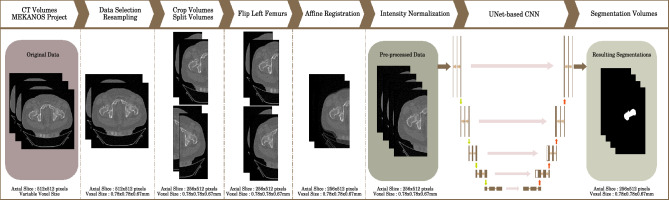
Bone segmentation is an important step to perform biomechanical failure load simulations on in-vivo CT data of patients with bone metastasis, as it is a mandatory operation to obtain meshes needed for numerical simulations. Segmentation can be a tedious and time consuming task when done manually, and expert segmentations are subject to intra- and inter-operator variability. Deep learning methods are increasingly employed to automatically carry out image segmentation tasks. These networks usually need to be trained on a large image dataset along with the manual segmentations to maximize generalization to new images, but it is not always possible to have access to a multitude of CT-scans with the associated ground truth. It then becomes necessary to use training techniques to make the best use of the limited available data. In this paper, we propose a dedicated pipeline of preprocessing, deep learning based segmentation method and post-processing for in-vivo human femurs and vertebrae segmentation from CT-scans volumes. We experimented with three U-Net architectures and showed that out-of-the-box models enable automatic and high-quality volume segmentation if carefully trained. We compared the failure load simulation results obtained on femurs and vertebrae using either automatic or manual segmentations and studied the sensitivity of the simulations on small variations of the automatic segmentation. The failure loads obtained using automatic segmentations were comparable to those obtained using manual expert segmentations for all the femurs and vertebrae tested, demonstrating the effectiveness of the automated segmentation approach for failure load simulations.
骨分割是对骨转移患者的体内CT数据进行生物力学破坏载荷模拟的重要步骤,因为它是获取数值模拟所需网格的必要操作。手动进行分割时,这可能是一项繁琐且耗时的任务,而且专家分割会受到操作者内部和操作者之间差异的影响。深度学习方法越来越多地用于自动执行图像分割任务。这些网络通常需要在大型图像数据集上结合手动分割进行训练,以最大程度地提高对新图像的泛化能力,但并非总是能够获得大量带有相关真实标注的CT扫描数据。因此,有必要使用训练技术来充分利用有限的可用数据。在本文中,我们提出了一种专门的预处理、基于深度学习的分割方法和后处理流程,用于从CT扫描体积中对体内人体股骨和椎骨进行分割。我们对三种U-Net架构进行了实验,结果表明,如果经过仔细训练,开箱即用的模型能够实现自动且高质量的体积分割。我们比较了使用自动分割和手动分割在股骨和椎骨上获得的破坏载荷模拟结果,并研究了模拟对自动分割小变化的敏感性。对于所有测试的股骨和椎骨,使用自动分割获得的破坏载荷与使用手动专家分割获得的破坏载荷相当,这证明了自动分割方法在破坏载荷模拟中的有效性。