Togni Lucrezia, Furlani Michele, Belloni Alessia, Riberti Nicole, Giuliani Alessandra, Notarstefano Valentina, Santoni Chiara, Giorgini Elisabetta, Rubini Corrado, Santarelli Andrea, Mascitti Marco
Department of Clinical Specialistic and Dental Sciences, Marche Polytechnic University, via Tronto 10, 60126 Ancona, Italy.
Department of Life and Environmental Science, Marche Polytechnic University, via Brecce Bianche, Ancona, Italy.
iScience. 2024 Jun 17;27(7):110303. doi: 10.1016/j.isci.2024.110303. eCollection 2024 Jul 19.
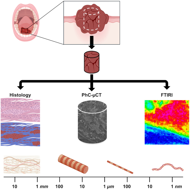
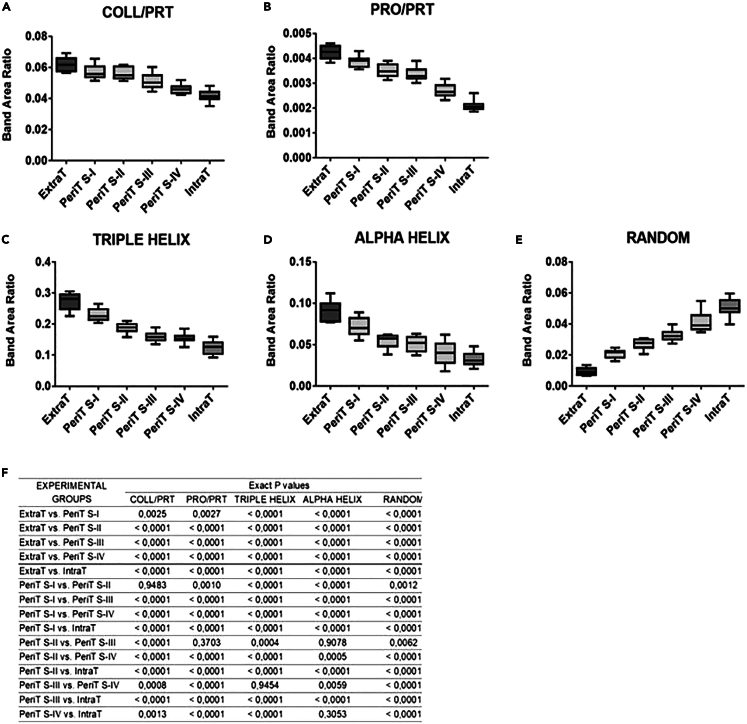
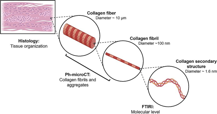
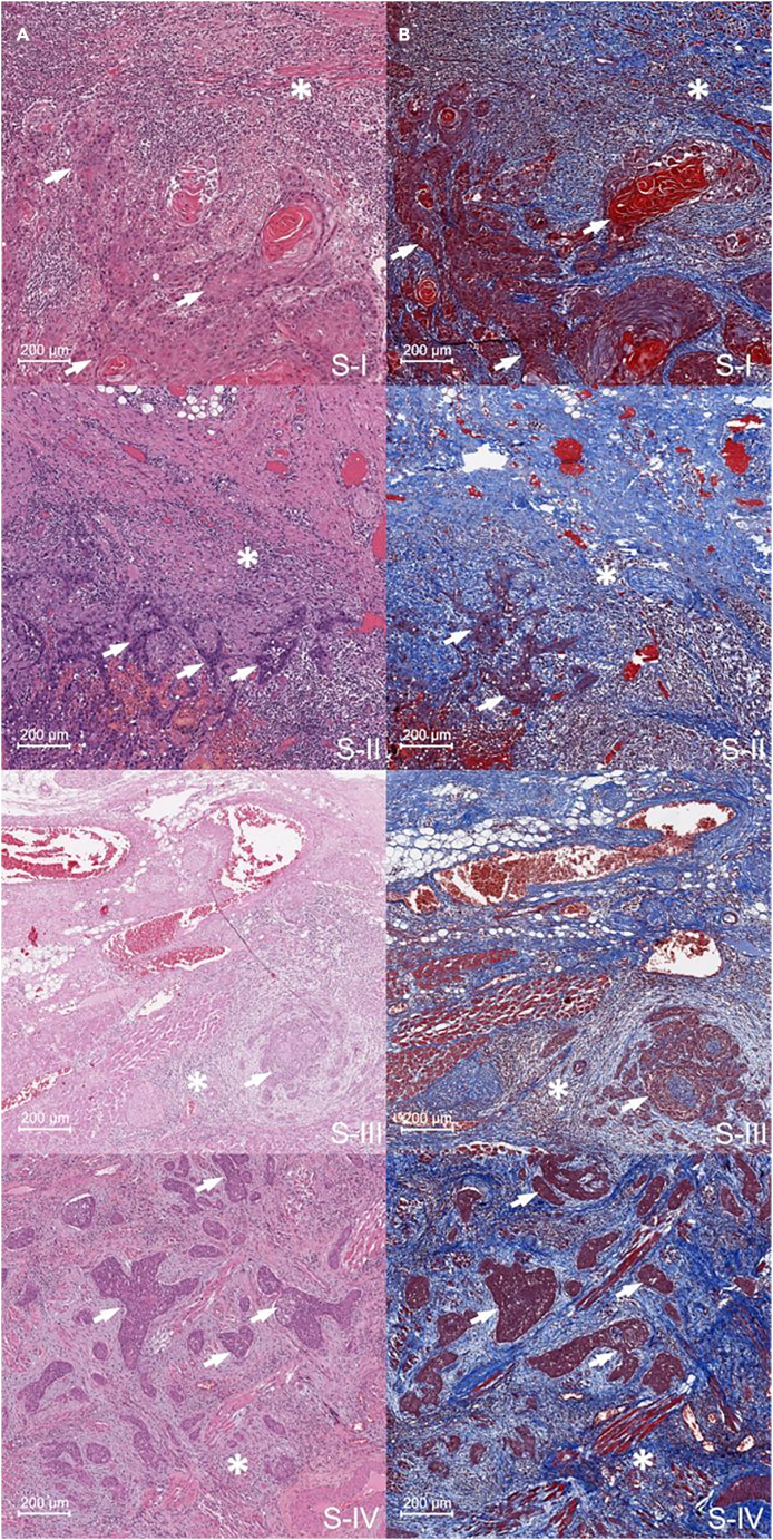
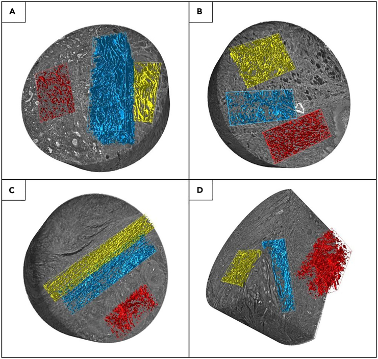
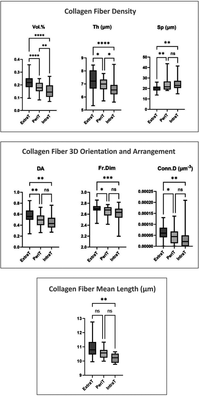
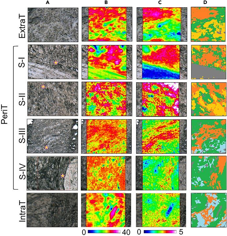
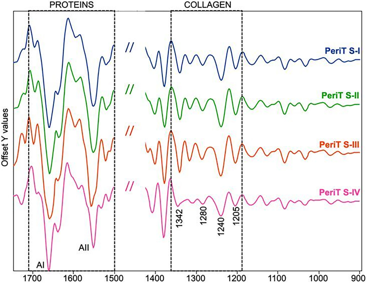
High resolution analysis of collagen bundles could provide information on tumor onset and evolution. This study was focused on the microarchitecture and biomolecular organization of collagen bundles in oral tongue squamous cell carcinoma (OTSCC). Thirty-five OTSCC biopsy samples were analyzed by synchrotron-based phase-contrast microcomputed tomography and Fourier transform infrared imaging (FTIRI) spectroscopy. PhC-microCT evidenced the presence of reduced and disorganized collagen in the tumor area compared to the extratumoral (ExtraT) one. FTIRI also revealed a reduction of folded secondary structures in the tumor area, and highlighted differences in the peritumoral (PeriT) areas in relation with the OTSCC stage, whereby a significantly lower amount of collagen with less organized fibers was found in the PeriT stroma of advanced-OTSCC stages. Interestingly, no significant morphometrical mismatches were detected in the same region by PhC-microCT analysis. These results suggest that biomolecular alterations in the OTSCC stroma temporally anticipate structural modifications of collagen bundle microarchitecture.
对胶原纤维束进行高分辨率分析可为肿瘤的发生和发展提供信息。本研究聚焦于口腔舌鳞状细胞癌(OTSCC)中胶原纤维束的微观结构和生物分子组织。通过基于同步辐射的相衬显微计算机断层扫描和傅里叶变换红外成像(FTIRI)光谱对35个OTSCC活检样本进行了分析。相衬显微计算机断层扫描(PhC-microCT)显示,与肿瘤外(ExtraT)区域相比,肿瘤区域的胶原减少且排列紊乱。FTIRI还揭示了肿瘤区域折叠二级结构的减少,并突出了肿瘤周围(PeriT)区域与OTSCC分期相关的差异,即在晚期OTSCC阶段的PeriT基质中发现胶原量显著减少且纤维组织较少。有趣的是,通过PhC-microCT分析在同一区域未检测到明显的形态计量学不匹配。这些结果表明,OTSCC基质中的生物分子改变在时间上先于胶原纤维束微观结构的结构改变。