• 文献检索
  • 文档翻译
  • 深度研究
  • 学术资讯
  • Suppr Zotero 插件Zotero 插件
  • 邀请有礼
  • 套餐&价格
  • 历史记录
应用&插件
Suppr Zotero 插件Zotero 插件浏览器插件Mac 客户端Windows 客户端微信小程序
定价
高级版会员购买积分包购买API积分包
服务
文献检索文档翻译深度研究API 文档MCP 服务
关于我们
关于 Suppr公司介绍联系我们用户协议隐私条款
关注我们

Suppr 超能文献

核心技术专利:CN118964589B侵权必究
粤ICP备2023148730 号-1Suppr @ 2026

文献检索

告别复杂PubMed语法,用中文像聊天一样搜索,搜遍4000万医学文献。AI智能推荐,让科研检索更轻松。

立即免费搜索

文件翻译

保留排版,准确专业,支持PDF/Word/PPT等文件格式,支持 12+语言互译。

免费翻译文档

深度研究

AI帮你快速写综述,25分钟生成高质量综述,智能提取关键信息,辅助科研写作。

立即免费体验

使用计算机断层扫描(CT)对一种市售的用于肺结节检测和特征分析的计算机辅助检测(CAD)系统进行验证。

Validation of a commercially available CAD-system for lung nodule detection and characterization using CT-scans.

作者信息

Paramasamy Jasika, Mandal Souvik, Blomjous Maurits, Mulders Ties, Bos Daniel, Aerts Joachim G J V, Vanapalli Prakash, Challa Vikash, Sathyamurthy Saigopal, Devi Ranjana, Jain Ritvik, Visser Jacob J

机构信息

Department of Radiology and Nuclear Medicine, Erasmus Medical Center, Dr. Molewaterplein 40, 3015 GD, Rotterdam, The Netherlands.

Qure.ai, Level 7, Oberoi Commerz II, Goregaon East, Mumbai, 400063, India.

出版信息

Eur Radiol. 2025 Feb;35(2):1076-1088. doi: 10.1007/s00330-024-10969-0. Epub 2024 Jul 23.

DOI:10.1007/s00330-024-10969-0
PMID:39042303
原文链接:https://pmc.ncbi.nlm.nih.gov/articles/PMC11782423/
Abstract

OBJECTIVES

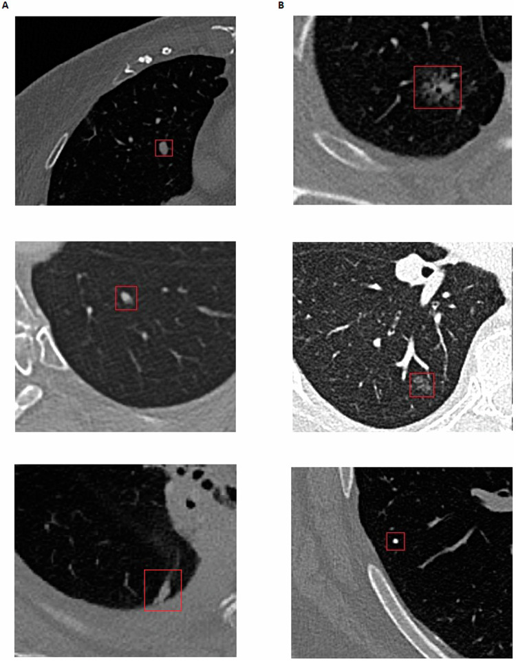
This study aims to externally validate a commercially available Computer-Aided Detection (CAD)-system for the automatic detection and characterization of solid, part-solid, and ground-glass lung nodules (LN) on CT scans.

METHODS

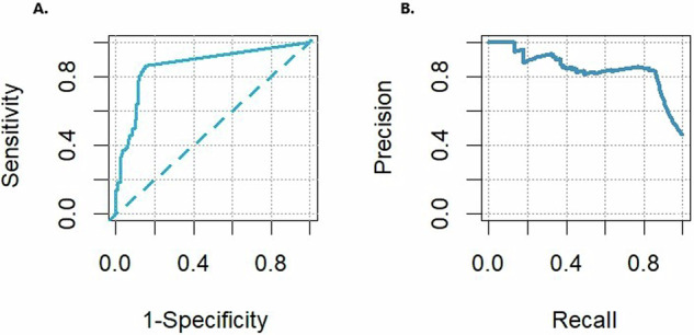
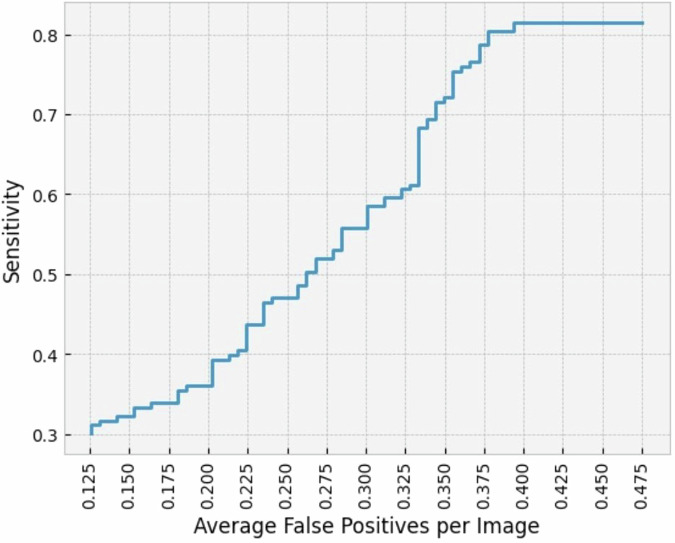
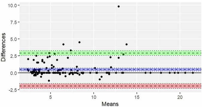
This retrospective study encompasses 263 chest CT scans performed between January 2020 and December 2021 at a Dutch university hospital. All scans were read by a radiologist (R1) and compared with the initial radiology report. Conflicting scans were assessed by an adjudicating radiologist (R2). All scans were also processed by CAD. The standalone performance of CAD in terms of sensitivity and false-positive (FP)-rate for detection was calculated together with the sensitivity for characterization, including texture, calcification, speculation, and location. The R1's detection sensitivity was also assessed.

RESULTS

A total of 183 true nodules were identified in 121 nodule-containing scans (142 non-nodule-containing scans), of which R1 identified 165/183 (90.2%). CAD detected 149 nodules, of which 12 were not identified by R1, achieving a sensitivity of 149/183 (81.4%) with an FP-rate of 49/121 (0.405). CAD's detection sensitivity for solid, part-solid, and ground-glass LNs was 82/94 (87.2%), 42/47 (89.4%), and 25/42 (59.5%), respectively. The classification accuracy for solid, part-solid, and ground-glass LNs was 81/82 (98.8%), 16/42 (38.1%), and 18/25 (72.0%), respectively. Additionally, CAD demonstrated overall classification accuracies of 137/149 (91.9%), 123/149 (82.6%), and 141/149 (94.6%) for calcification, spiculation, and location, respectively.

CONCLUSIONS

Although the overall detection rate of this system slightly lags behind that of a radiologist, CAD is capable of detecting different LNs and thereby has the potential to enhance a reader's detection rate. While promising characterization performances are obtained, the tool's performance in terms of texture classification remains a subject of concern.

CLINICAL RELEVANCE STATEMENT

Numerous lung nodule computer-aided detection-systems are commercially available, with some of them solely being externally validated based on their detection performance on solid nodules. We encourage researchers to assess performances by incorporating all relevant characteristics, including part-solid and ground-glass nodules.

KEY POINTS

Few computer-aided detection (CAD) systems are externally validated for automatic detection and characterization of lung nodules. A detection sensitivity of 81.4% and an overall texture classification sensitivity of 77.2% were measured utilizing CAD. CAD has the potential to increase single reader detection rate, however, improvement in texture classification is required.

摘要

目的

本研究旨在对一种商用计算机辅助检测(CAD)系统进行外部验证,该系统用于在CT扫描中自动检测和鉴别实性、部分实性和磨玻璃肺结节(LN)。

方法

这项回顾性研究涵盖了2020年1月至2021年12月在一家荷兰大学医院进行的263例胸部CT扫描。所有扫描均由一名放射科医生(R1)阅片,并与初始放射学报告进行比较。存在争议的扫描由一名裁决放射科医生(R2)评估。所有扫描也由CAD进行处理。计算CAD在检测方面的灵敏度和假阳性(FP)率的独立性能,以及鉴别方面的灵敏度,包括纹理、钙化、毛刺和位置。同时评估R1的检测灵敏度。

结果

在121例含有结节的扫描(142例不含结节的扫描)中总共识别出183个真结节,其中R1识别出165/183(90.2%)。CAD检测出149个结节,其中12个未被R1识别,灵敏度达到149/183(81.4%),FP率为49/121(0.405)。CAD对实性、部分实性和磨玻璃LN的检测灵敏度分别为82/94(87.2%)、42/47(89.4%)和25/42(59.5%)。实性、部分实性和磨玻璃LN的分类准确率分别为81/82(98.8%)、16/42(38.1%)和18/25(72.0%)。此外,CAD在钙化、毛刺和位置方面的总体分类准确率分别为137/149(91.9%)、123/149(82.6%)和141/149(94.6%)。

结论

尽管该系统的总体检测率略落后于放射科医生,但CAD能够检测不同的LN,因此有潜力提高阅片者的检测率。虽然获得了有前景的鉴别性能,但该工具在纹理分类方面的性能仍是一个值得关注的问题。

临床相关性声明

有许多商用肺结节计算机辅助检测系统,其中一些仅基于其对实性结节的检测性能进行了外部验证。我们鼓励研究人员通过纳入所有相关特征,包括部分实性和磨玻璃结节,来评估性能。

关键点

很少有计算机辅助检测(CAD)系统针对肺结节的自动检测和鉴别进行外部验证。使用CAD测量的检测灵敏度为81.4%,总体纹理分类灵敏度为77.2%。CAD有潜力提高单阅片者的检测率,然而,纹理分类方面需要改进。

https://cdn.ncbi.nlm.nih.gov/pmc/blobs/6512/11782423/c8d0eb9f856d/330_2024_10969_Fig6_HTML.jpg
https://cdn.ncbi.nlm.nih.gov/pmc/blobs/6512/11782423/2dd059ab545a/330_2024_10969_Fig1_HTML.jpg
https://cdn.ncbi.nlm.nih.gov/pmc/blobs/6512/11782423/2f70c8768549/330_2024_10969_Fig2_HTML.jpg
https://cdn.ncbi.nlm.nih.gov/pmc/blobs/6512/11782423/c06229774883/330_2024_10969_Fig3_HTML.jpg
https://cdn.ncbi.nlm.nih.gov/pmc/blobs/6512/11782423/9f5bc27d1edf/330_2024_10969_Fig4_HTML.jpg
https://cdn.ncbi.nlm.nih.gov/pmc/blobs/6512/11782423/d1142f1e674c/330_2024_10969_Fig5_HTML.jpg
https://cdn.ncbi.nlm.nih.gov/pmc/blobs/6512/11782423/c8d0eb9f856d/330_2024_10969_Fig6_HTML.jpg

相似文献

1
Validation of a commercially available CAD-system for lung nodule detection and characterization using CT-scans.使用计算机断层扫描(CT)对一种市售的用于肺结节检测和特征分析的计算机辅助检测(CAD)系统进行验证。
Eur Radiol. 2025 Feb;35(2):1076-1088. doi: 10.1007/s00330-024-10969-0. Epub 2024 Jul 23.
2
Validation of a deep learning computer aided system for CT based lung nodule detection, classification, and growth rate estimation in a routine clinical population.深度学习计算机辅助系统对常规临床人群 CT 肺结节检测、分类和生长速度评估的验证。
PLoS One. 2022 May 5;17(5):e0266799. doi: 10.1371/journal.pone.0266799. eCollection 2022.
3
Computer-aided diagnosis (CAD) of subsolid nodules: Evaluation of a commercial CAD system.亚实性结节的计算机辅助诊断(CAD):对一种商用CAD系统的评估
Eur J Radiol. 2016 Oct;85(10):1728-1734. doi: 10.1016/j.ejrad.2016.07.011. Epub 2016 Jul 18.
4
Commercially available computer-aided detection system for pulmonary nodules on thin-section images using 64 detectors-row CT: preliminary study of 48 cases.使用64排探测器CT对薄层图像上肺结节进行商用计算机辅助检测系统:48例初步研究
Acad Radiol. 2009 Aug;16(8):924-33. doi: 10.1016/j.acra.2009.01.030. Epub 2009 Apr 25.
5
Computer-aided detection of pulmonary nodules: a comparative study using the public LIDC/IDRI database.肺结节的计算机辅助检测:使用公共LIDC/IDRI数据库的对比研究。
Eur Radiol. 2016 Jul;26(7):2139-47. doi: 10.1007/s00330-015-4030-7. Epub 2015 Oct 6.
6
A cloud-based computer-aided detection system improves identification of lung nodules on computed tomography scans of patients with extra-thoracic malignancies.基于云的计算机辅助检测系统可提高对伴有胸外恶性肿瘤的患者 CT 扫描中肺结节的识别能力。
Eur Radiol. 2019 Jan;29(1):144-152. doi: 10.1007/s00330-018-5528-6. Epub 2018 Jun 15.
7
Benefit of computer-aided detection analysis for the detection of subsolid and solid lung nodules on thin- and thick-section CT.计算机辅助检测分析对薄层和厚层 CT 上亚实性和实性肺结节检测的益处。
AJR Am J Roentgenol. 2013 Jan;200(1):74-83. doi: 10.2214/AJR.11.7532.
8
External validation of the performance of commercially available deep-learning-based lung nodule detection on low-dose CT images for lung cancer screening in Japan.基于深度学习的商用肺癌结节检测在日本低剂量CT图像上用于肺癌筛查的性能外部验证。
Jpn J Radiol. 2025 Apr;43(4):634-640. doi: 10.1007/s11604-024-01704-2. Epub 2024 Nov 30.
9
Performance of Lung Nodule Computer-Aided Detection Systems on Standard-Dose and Low-Dose Pediatric CT Scans: An Intraindividual Comparison.肺结节计算机辅助检测系统在标准剂量和低剂量儿科CT扫描上的性能:个体内比较
AJR Am J Roentgenol. 2025 Jan;224(1):e2431972. doi: 10.2214/AJR.24.31972. Epub 2024 Oct 9.
10
Solid, part-solid, or non-solid?: classification of pulmonary nodules in low-dose chest computed tomography by a computer-aided diagnosis system.实性、部分实性或非实性?:计算机辅助诊断系统在低剂量胸部 CT 中对肺结节的分类。
Invest Radiol. 2015 Mar;50(3):168-73. doi: 10.1097/RLI.0000000000000121.

引用本文的文献

1
Cross-validation of an artificial intelligence tool for fracture classification and localization on conventional radiography in Dutch population.荷兰人群中用于传统X线摄影骨折分类和定位的人工智能工具的交叉验证
Insights Imaging. 2025 Jul 3;16(1):150. doi: 10.1186/s13244-025-02034-1.
2
Computer-aided Nodule Detection in the Lung Apices in Head and Neck Computed Tomography Angiography: An Unexpected Opportunity.头颈部计算机断层扫描血管造影中肺尖部的计算机辅助结节检测:一个意外的机遇。
J Thorac Imaging. 2025 Sep 1;40(5):e0836. doi: 10.1097/RTI.0000000000000836.
3
China Protocol for early screening, precise diagnosis, and individualized treatment of lung cancer.

本文引用的文献

1
Radiology AI Deployment and Assessment Rubric (RADAR) to bring value-based AI into radiological practice.放射学人工智能部署与评估准则(RADAR),将基于价值的人工智能引入放射学实践。
Insights Imaging. 2024 Feb 5;15(1):34. doi: 10.1186/s13244-023-01599-z.
2
The Growing Problem of Radiologist Shortages: Korean Perspective.放射科医生短缺问题日益严重:韩国视角
Korean J Radiol. 2023 Dec;24(12):1173-1175. doi: 10.3348/kjr.2023.1010.
3
Trends in the incidence of pulmonary nodules in chest computed tomography: 10-year results from two Dutch hospitals.
中国肺癌早期筛查、精准诊断及个体化治疗方案
Signal Transduct Target Ther. 2025 May 27;10(1):175. doi: 10.1038/s41392-025-02256-1.
胸部 CT 检出肺结节的发病趋势:来自荷兰两家医院的 10 年研究结果。
Eur Radiol. 2023 Nov;33(11):8279-8288. doi: 10.1007/s00330-023-09826-3. Epub 2023 Jun 20.
4
Intra- and Inter-Reader Variations in Lung Nodule Measurements: Influences of Nodule Size, Location, and Observers.肺结节测量中阅片者内部和阅片者间的差异:结节大小、位置及观察者的影响
Diagnostics (Basel). 2022 Sep 26;12(10):2319. doi: 10.3390/diagnostics12102319.
5
External Validation of Deep Learning Algorithms for Radiologic Diagnosis: A Systematic Review.用于放射诊断的深度学习算法的外部验证:一项系统评价。
Radiol Artif Intell. 2022 May 4;4(3):e210064. doi: 10.1148/ryai.210064. eCollection 2022 May.
6
Validation of a deep learning computer aided system for CT based lung nodule detection, classification, and growth rate estimation in a routine clinical population.深度学习计算机辅助系统对常规临床人群 CT 肺结节检测、分类和生长速度评估的验证。
PLoS One. 2022 May 5;17(5):e0266799. doi: 10.1371/journal.pone.0266799. eCollection 2022.
7
Evaluating the Patient With a Pulmonary Nodule: A Review.评估肺部结节患者:综述。
JAMA. 2022 Jan 18;327(3):264-273. doi: 10.1001/jama.2021.24287.
8
Performance of a deep learning-based lung nodule detection system as an alternative reader in a Chinese lung cancer screening program.深度学习辅助肺结节检测系统作为中国肺癌筛查项目中替代读片者的性能。
Eur J Radiol. 2022 Jan;146:110068. doi: 10.1016/j.ejrad.2021.110068. Epub 2021 Nov 24.
9
Seasonal prevalence and characteristics of low-dose CT detected lung nodules in a general Dutch population.荷兰一般人群中低剂量 CT 检测到的肺结节的季节性流行率和特征。
Sci Rep. 2021 Apr 28;11(1):9139. doi: 10.1038/s41598-021-88328-y.
10
Screening for Lung Cancer: US Preventive Services Task Force Recommendation Statement.肺癌筛查:美国预防服务工作组推荐声明。
JAMA. 2021 Mar 9;325(10):962-970. doi: 10.1001/jama.2021.1117.