• 文献检索
  • 文档翻译
  • 深度研究
  • 学术资讯
  • Suppr Zotero 插件Zotero 插件
  • 邀请有礼
  • 套餐&价格
  • 历史记录
应用&插件
Suppr Zotero 插件Zotero 插件浏览器插件Mac 客户端Windows 客户端微信小程序
定价
高级版会员购买积分包购买API积分包
服务
文献检索文档翻译深度研究API 文档MCP 服务
关于我们
关于 Suppr公司介绍联系我们用户协议隐私条款
关注我们

Suppr 超能文献

核心技术专利:CN118964589B侵权必究
粤ICP备2023148730 号-1Suppr @ 2026

文献检索

告别复杂PubMed语法,用中文像聊天一样搜索,搜遍4000万医学文献。AI智能推荐,让科研检索更轻松。

立即免费搜索

文件翻译

保留排版,准确专业,支持PDF/Word/PPT等文件格式,支持 12+语言互译。

免费翻译文档

深度研究

AI帮你快速写综述,25分钟生成高质量综述,智能提取关键信息,辅助科研写作。

立即免费体验

胎儿超声皮质发育与异常:系统评价与荟萃分析

Sonographic Cortical Development and Anomalies in the Fetus: A Systematic Review and Meta-Analysis.

作者信息

Mappa Ilenia, Di Mascio Daniele, Carbone Luigi, Lu Jia Li Angela, Sorrenti Sara, Patelli Chiara, D'Amico Alice, Matarrelli Barbara, Giuliani Giulia Andrea, Neola Daniele, Di Girolamo Raffaella, Sarno Laura, Khalil Asma, Rizzo Giuseppe, Maruotti Giuseppe Maria, D'Antonio Francesco

机构信息

Department of Obstetrics and Gynecology, Fondazione Policlinico Tor Vergata, University of Rome Tor Vergata, 00133 Rome, Italy.

Department of Maternal and Child Health and Urological Sciences, Sapienza University of Rome, 00161 Roma, Italy.

出版信息

Biomedicines. 2024 Jun 24;12(7):1397. doi: 10.3390/biomedicines12071397.

DOI:10.3390/biomedicines12071397
PMID:39061971
原文链接:https://pmc.ncbi.nlm.nih.gov/articles/PMC11273757/
Abstract

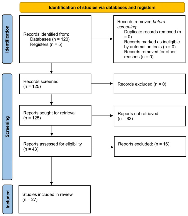
The aim of this systematic review is to report the normal cortical development of different fetal cerebral fissures on ultrasound, describe associated anomalies in fetuses with cortical malformations, and evaluate the quality of published charts of cortical fissures. The inclusion criteria were studies reporting development, anomalies, and reference charts of fetal cortical structures on ultrasound. The outcomes observed were the timing of the appearance of different cortical fissures according to different gestational age windows, associated central nervous system (CNS) and extra-CNS anomalies detected at ultrasound in fetuses with cortical malformation, and rate of fetuses with isolated anomaly. Furthermore, we performed a critical evaluation of the published reference charts for cortical development on ultrasound. Random-effect meta-analyses of proportions were used to combine the data. Twenty-seven studies (6875 fetuses) were included. Sylvian fissure was visualized on ultrasound in 97.69% (95% CI 92.0-100) of cases at 18-19, 98.17% (95% CI 94.8-99.8) at 20-21, 98.94% (95% CI 97.0-99.9) at 22-23, and in all cases from 24 weeks of gestation. Parieto-occipital fissure was visualized in 81.56% (95% CI 48.4-99.3) of cases at 18-19, 96.59% (95% CI 83.2-99.8) at 20-21, 96.85% (95% CI 88.8-100) at 22-23, and in all cases from 24 weeks of gestation, while the corresponding figures for calcarine fissure were 37.27% (95% CI 0.5-89.6), 80.42% (95% CI 50.2-98.2), 89.18% (95% CI 74.0-98.2), and 96.02% (95% CI 96.9-100). Malformations of cortical development were diagnosed as an isolated finding at ultrasound in 6.21% (95% CI 2.9-10.9) of cases, while they were associated with additional CNS anomalies in 93.79% (95% CI 89.1-97.2) of cases. These findings highlight the need for large studies specifically looking at the timing of the appearance of the different brain sulci. Standardized algorithms for prenatal assessment of fetuses at high risk of malformations of cortical development are also warranted.

摘要

本系统评价的目的是报告超声检查中不同胎儿脑沟的正常皮质发育情况,描述皮质发育畸形胎儿的相关异常,并评估已发表的皮质沟图表的质量。纳入标准为报告超声检查中胎儿皮质结构发育、异常及参考图表的研究。观察到的结果包括不同孕周窗口下不同皮质沟出现的时间、皮质发育畸形胎儿超声检查中检测到的相关中枢神经系统(CNS)和中枢神经系统外异常,以及孤立异常胎儿的比例。此外,我们对已发表的超声皮质发育参考图表进行了批判性评估。采用随机效应meta分析比例法合并数据。纳入了27项研究(6875例胎儿)。在妊娠18 - 19周时,97.69%(95%CI 92.0 - 100)的病例超声可显示外侧裂;20 - 21周时为98.17%(95%CI 94.8 - 99.8);22 - 23周时为98.94%(95%CI 97.0 - 99.9);妊娠24周及以后的所有病例均可显示。在妊娠18 - 19周时,81.56%(95%CI 48.4 - 99.3)的病例超声可显示顶枕裂;20 - 21周时为96.59%(95%CI 83.2 - 99.8);22 - 23周时为96.85%(95%CI 88.8 - 100);妊娠24周及以后的所有病例均可显示。而距状沟的相应数据分别为37.27%(95%CI 0.5 - 89.6)、80.42%(95%CI 50.2 - 98.2)、89.18%(95%CI 74.0 - 98.2)和96.02%(95%CI 96.9 - 100)。超声检查中,6.21%(95%CI 2.9 - 10.9)的病例皮质发育畸形被诊断为孤立性发现,而93.79%(95%CI 89.1 - 97.2)的病例与其他中枢神经系统异常相关。这些发现凸显了开展专门研究不同脑沟出现时间的大型研究的必要性。制定针对皮质发育畸形高风险胎儿的产前评估标准化算法也很有必要。

https://cdn.ncbi.nlm.nih.gov/pmc/blobs/d570/11273757/e63f20abe232/biomedicines-12-01397-g001.jpg

相似文献

1
Sonographic Cortical Development and Anomalies in the Fetus: A Systematic Review and Meta-Analysis.胎儿超声皮质发育与异常:系统评价与荟萃分析
Biomedicines. 2024 Jun 24;12(7):1397. doi: 10.3390/biomedicines12071397.
2
Role of magnetic resonance imaging in fetuses with mild or moderate ventriculomegaly in the era of fetal neurosonography: systematic review and meta-analysis.磁共振成像在胎儿神经超声时代轻度或中度脑室扩张胎儿中的作用:系统评价和荟萃分析。
Ultrasound Obstet Gynecol. 2019 Aug;54(2):164-171. doi: 10.1002/uog.20197. Epub 2019 Jul 11.
3
Increased Sylvian fissure angle as early sonographic sign of malformation of cortical development.大脑皮质发育畸形的早期超声征象:大脑外侧裂角度增大。
Ultrasound Obstet Gynecol. 2019 Aug;54(2):199-206. doi: 10.1002/uog.20171. Epub 2019 Jul 8.
4
Role of prenatal magnetic resonance imaging in fetuses with isolated anomalies of corpus callosum: multinational study.孤立性胼胝体发育不全胎儿的产前磁共振成像作用:多国研究。
Ultrasound Obstet Gynecol. 2021 Jul;58(1):26-33. doi: 10.1002/uog.23612.
5
Prenatal ultrasound findings of lissencephaly associated with Miller-Dieker syndrome and comparison with pre- and postnatal magnetic resonance imaging.与米勒-迪克尔综合征相关的无脑回畸形的产前超声检查结果及与产前和产后磁共振成像的比较
Ultrasound Obstet Gynecol. 2004 Dec;24(7):716-23. doi: 10.1002/uog.1777.
6
Role of prenatal magnetic resonance imaging in fetuses with isolated mild or moderate ventriculomegaly in the era of neurosonography: international multicenter study.产前磁共振成像在神经超声时代孤立性轻度或中度脑室扩张胎儿中的作用:国际多中心研究。
Ultrasound Obstet Gynecol. 2020 Sep;56(3):340-347. doi: 10.1002/uog.21974.
7
Role of prenatal magnetic resonance imaging in fetuses with isolated severe ventriculomegaly at neurosonography: A multicenter study.产前磁共振成像在神经超声检查孤立性重度脑室扩张胎儿中的作用:一项多中心研究。
Eur J Obstet Gynecol Reprod Biol. 2021 Dec;267:105-110. doi: 10.1016/j.ejogrb.2021.10.014. Epub 2021 Oct 23.
8
Diagnosis of fetal cortical abnormalities by new reference charts for assessment of sylvian fissure biometry.通过新的大脑外侧裂生物测量参考图表诊断胎儿皮质异常。
Prenat Diagn. 2023 Jul;43(8):1066-1078. doi: 10.1002/pd.6359. Epub 2023 May 15.
9
Transvaginal three-dimensional ultrasound assessment of Sylvian fissures at 18-30 weeks' gestation.经阴道三维超声评估 18-30 孕周大脑外侧裂。
Ultrasound Obstet Gynecol. 2019 Aug;54(2):190-198. doi: 10.1002/uog.20172. Epub 2019 Jul 8.
10
Role of fetal magnetic resonance imaging in fetuses with congenital cytomegalovirus infection: multicenter study.先天性巨细胞病毒感染胎儿的胎儿磁共振成像作用:多中心研究。
Ultrasound Obstet Gynecol. 2023 Jan;61(1):67-73. doi: 10.1002/uog.26054.

引用本文的文献

1
Counselling in Fetal Medicine: Uncomplicated Twin Pregnancies.胎儿医学中的咨询:单绒双羊双胎妊娠
J Clin Med. 2024 Dec 3;13(23):7355. doi: 10.3390/jcm13237355.
2
Counselling in Fetal Medicine: Complications of Monochorionic Diamniotic Twin Pregnancies.胎儿医学中的咨询:单绒毛膜双羊膜囊双胎妊娠的并发症
J Clin Med. 2024 Nov 30;13(23):7295. doi: 10.3390/jcm13237295.

本文引用的文献

1
Modeling fetal cortical development by quantile regression for gestational age and head circumference: a prospective cross sectional study.通过对孕龄和头围进行分位数回归来对胎儿皮质发育进行建模:一项前瞻性的横断面研究。
J Perinat Med. 2023 Aug 21;51(9):1212-1219. doi: 10.1515/jpm-2023-0176. Print 2023 Nov 27.
2
Prenatal diagnosis of microcephaly with simplified gyral pattern: series of eight cases.简化脑回模式小头畸形的产前诊断:8 例系列。
Ultrasound Obstet Gynecol. 2024 Feb;63(2):271-275. doi: 10.1002/uog.27450. Epub 2024 Jan 11.
3
A New Parameter to Evaluate Fetal Sylvian Fissure by Transabdominal 2-D Ultrasound.
经腹二维超声评估胎儿侧裂池的新参数。
Ultrasound Med Biol. 2023 Sep;49(9):2054-2059. doi: 10.1016/j.ultrasmedbio.2023.05.010. Epub 2023 Jun 10.
4
Diagnosis of fetal cortical abnormalities by new reference charts for assessment of sylvian fissure biometry.通过新的大脑外侧裂生物测量参考图表诊断胎儿皮质异常。
Prenat Diagn. 2023 Jul;43(8):1066-1078. doi: 10.1002/pd.6359. Epub 2023 May 15.
5
Corpus callosal reference ranges: systematic review of methodology of biometric chart construction and measurements obtained.胼胝体参考范围:生物测量图表构建和获得的测量方法的系统评价。
Ultrasound Obstet Gynecol. 2023 Aug;62(2):175-184. doi: 10.1002/uog.26187.
6
Malformation of cortical development with abnormal cortex: early ultrasound diagnosis between 14 and 24 weeks of gestation.皮质发育畸形伴皮质异常:14-24 孕周的早期超声诊断。
Ultrasound Obstet Gynecol. 2023 May;61(5):559-565. doi: 10.1002/uog.26139. Epub 2023 Apr 7.
7
Three-Dimensional Crystal Vue Imaging technology assessment of Sylvian fissures at 20-32+6 weeks' normal gestation.20至32⁺⁶周正常妊娠时大脑外侧裂的三维晶体视野成像技术评估
Eur Radiol. 2023 Apr;33(4):2358-2366. doi: 10.1007/s00330-022-09253-w. Epub 2022 Nov 17.
8
Role of fetal magnetic resonance imaging in fetuses with congenital cytomegalovirus infection: multicenter study.先天性巨细胞病毒感染胎儿的胎儿磁共振成像作用:多中心研究。
Ultrasound Obstet Gynecol. 2023 Jan;61(1):67-73. doi: 10.1002/uog.26054.
9
Methodological Quality of Fetal Brain Structure Charts for Screening Examination and Targeted Neurosonography: A Systematic Review.用于筛查检查和针对性神经超声检查的胎儿脑结构图表的方法学质量:一项系统评价。
Fetal Diagn Ther. 2022;49(4):145-158. doi: 10.1159/000521421. Epub 2022 Apr 22.
10
Role of Ultrasonography in the Evaluation of Normal Developmental Pattern of Fetal Cerebral Sulci Between 18 and 32 Weeks of Gestational Age.超声检查在评估孕18至32周胎儿脑沟正常发育模式中的作用。
Cureus. 2022 Feb 24;14(2):e22581. doi: 10.7759/cureus.22581. eCollection 2022 Feb.