Shbani Abdulrahman, Suleiman Qamar, Suleiman Fadi
Tartous university, Faculty of Medicine, Tartous, Syrian Arab Republic.
Tartous university, Faculty of Medicine, Tartous, Syrian Arab Republic.
Int J Surg Case Rep. 2024 Sep;122:110052. doi: 10.1016/j.ijscr.2024.110052. Epub 2024 Jul 20.
Retrorectal tumors are rare growths with various types which are found in the space behind the rectum. They can be diverse and are often diagnosed through imaging and surgery.
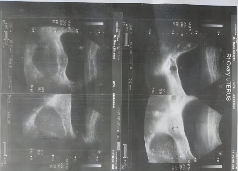
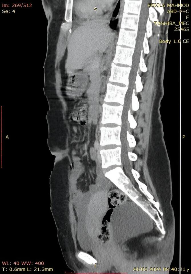
A 31-year-old female patient came to the clinic with concerns about irregular periods and constipation, but no history of abdominal pain, pelvic pressure, or weight loss. She had a previous surgery to remove an ovarian teratoma when she was three months old. Physical exams and lab tests showed no significant findings, except for a pelvic ultrasound that revealed a normal right ovary with a small follicle and a missing left ovary due to prior surgery for a dermoid cyst. Another cyst, measuring 8.2 × 9.3 × 5.7 cm, was found behind the uterus, believed to be a presacral cyst possibly originating from elsewhere. Further investigation with a CT scan confirmed the presence of a large cyst near the rectum, leading to an open surgical procedure to remove it. The cyst, located deep behind the rectum and next to the levator ani muscle, contained a substance resembling cheese with hair, suggesting a benign dermoid cyst with granulation tissue. The surgery was successful, and the diagnosis was confirmed through histopathological analysis.
Retrorectal teratomas are rare germ cell tumors that mainly affect children, often presenting with vague symptoms like constipation. Diagnosis involves imaging tests like ultrasound, CT scans, and MRI, with surgical removal being the primary treatment option. Recurrence rates are low with complete excision of benign tumors.
Retrorectal or presacral teratomas are rare tumors with vague symptoms, making diagnosis difficult. They are often detected late and require radiological assessment for surgical planning. Treatment success hinges on a coordinated effort by skilled radiologists and surgeons specializing in pelvic and oncological care to ensure favorable outcomes with lower recurrence rates and risks.
直肠后肿瘤是发生于直肠后方间隙的罕见肿瘤,类型多样。它们表现各异,常通过影像学检查和手术进行诊断。
一名31岁女性患者因月经不规律和便秘前来就诊,无腹痛、盆腔压迫感或体重减轻史。她在3个月大时曾接受过卵巢畸胎瘤切除术。体格检查和实验室检查均无明显异常,盆腔超声显示右侧卵巢正常,有一个小卵泡,左侧卵巢因既往皮样囊肿手术缺失。在子宫后方发现另一个囊肿,大小为8.2×9.3×5.7厘米,据信是一个可能起源于其他部位的骶前囊肿。CT扫描进一步检查证实直肠附近存在一个大囊肿,遂进行开放手术将其切除。该囊肿位于直肠后方深处,毗邻肛提肌,内含类似奶酪并伴有毛发的物质,提示为伴有肉芽组织的良性皮样囊肿。手术成功,通过组织病理学分析确诊。
直肠后畸胎瘤是罕见的生殖细胞肿瘤,主要影响儿童,常表现为便秘等模糊症状。诊断需要超声、CT扫描和MRI等影像学检查,手术切除是主要治疗选择。良性肿瘤完全切除后的复发率较低。
直肠后或骶前畸胎瘤是罕见肿瘤,症状模糊,诊断困难。它们常被发现较晚,手术规划需要进行放射学评估。治疗成功取决于专业的放射科医生和擅长盆腔及肿瘤护理的外科医生的协同努力,以确保获得较低复发率和风险的良好治疗效果。