Rendon C Javier, Sempere Lorenzo, Lauver Adam, Watts Stephanie W, Contreras G Andres
Department of Large Animal Clinical Sciences, College of Veterinary Medicine, Michigan State University, East Lansing, MI, United States.
Department of Radiology and Precision Health Program, Michigan State University, East Lansing, MI, United States.
Front Physiol. 2024 Jul 12;15:1411218. doi: 10.3389/fphys.2024.1411218. eCollection 2024.
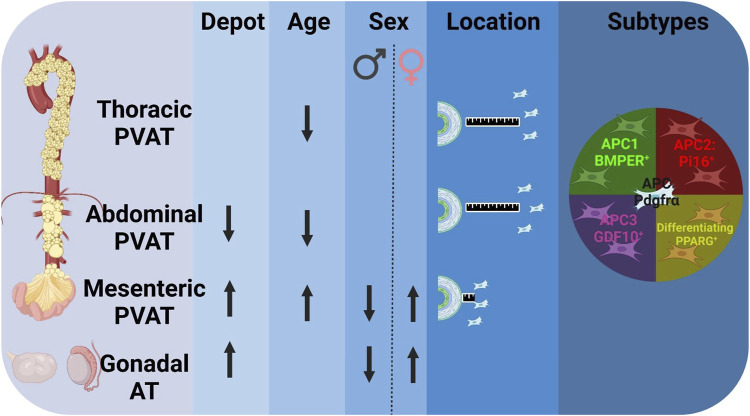
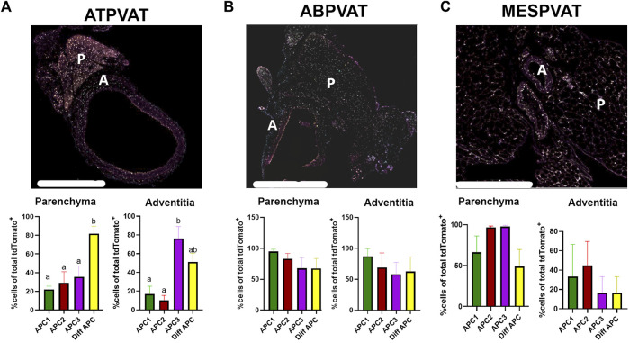
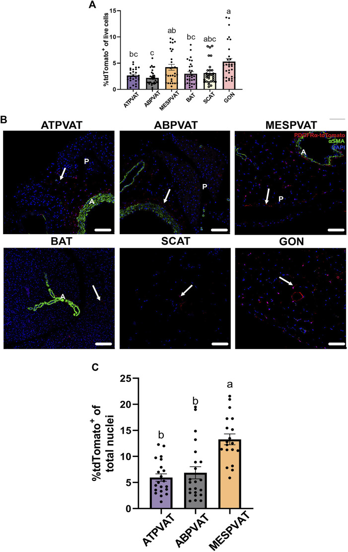
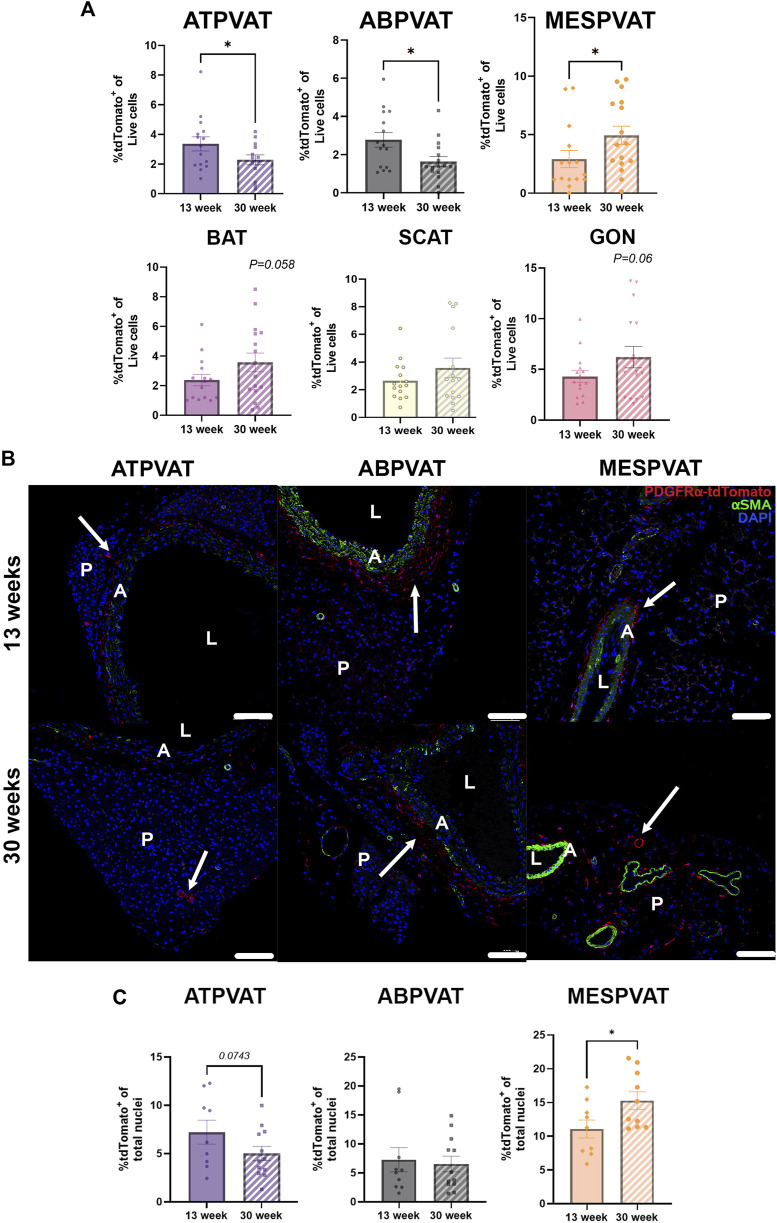
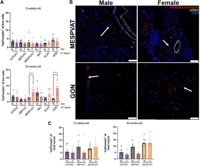
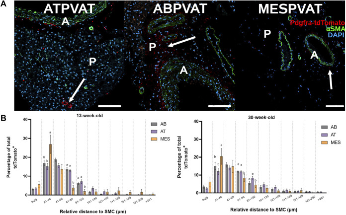
Perivascular adipose tissue (PVAT) regulates vascular function due to its capacity to synthesize vasoactive products and its mechanical properties. PVATs most abundant cells are adipocytes, and their populations are maintained by the maturation of adipocyte progenitor cells (APC), which may play a pivotal role in the pathogenesis of cardiovascular diseases. However, the distribution of APC within PVAT depots, their potential variation in spatial location, and the influence of sex and age on their abundance remain unknown. We hypothesize that APC abundance in PVAT is affected by location, age, sex and that APC subtypes have specific spatial distributions. PVAT from thoracic and abdominal aorta, and mesenteric arteries, and AT from interscapular, gonadal, and subcutaneous depots from 13-week and 30-week-old females and males Pdgfrα-CreERT2 x LSL-tdTomato mice (n = 28) were analyzed. Abdominal aorta PVAT had fewer progenitors than mesenteric PVAT and gonadal AT. Aging reduced the abundance of APC in the thoracic aorta but increased their numbers in mesenteric PVAT. Females had more APC than males in mesenteric PVAT and gonadal AT depots. APC exhibited unique spatial distribution in the aorta and mesenteric PVAT where they localized neighboring vasa vasorum and arteries. APC subtypes (APC1, APC2, APC3, diff APC) were identified in all PVAT depots. Thoracic aorta PVAT APC3 were located in the adventitia while diff APC were in the parenchyma. This study identified variability in APC populations based on depot, age, and sex. The distinctive spatial distribution and the presence of diverse APC subtypes suggest that they may contribute differently to cardiovascular diseases-induced PVAT remodeling.
血管周围脂肪组织(PVAT)因其合成血管活性产物的能力及其机械特性而调节血管功能。PVAT中最丰富的细胞是脂肪细胞,其数量通过脂肪细胞祖细胞(APC)的成熟得以维持,而APC可能在心血管疾病的发病机制中起关键作用。然而,APC在PVAT库中的分布、其空间位置的潜在变化以及性别和年龄对其丰度的影响仍然未知。我们假设PVAT中APC的丰度受位置、年龄、性别的影响,并且APC亚型具有特定的空间分布。对13周龄和30周龄的雌性和雄性Pdgfrα-CreERT2 x LSL-tdTomato小鼠(n = 28)的胸主动脉、腹主动脉和肠系膜动脉的PVAT,以及肩胛间、性腺和皮下库的脂肪组织(AT)进行了分析。腹主动脉PVAT中的祖细胞比肠系膜PVAT和性腺AT中的少。衰老减少了胸主动脉中APC的丰度,但增加了肠系膜PVAT中APC的数量。在肠系膜PVAT和性腺AT库中,雌性的APC比雄性多。APC在主动脉和肠系膜PVAT中表现出独特的空间分布,它们定位于邻近的血管滋养血管和动脉。在所有PVAT库中都鉴定出了APC亚型(APC1、APC2、APC3、diff APC)。胸主动脉PVAT中的APC3位于外膜,而diff APC位于实质。本研究确定了基于库、年龄和性别的APC群体的变异性。独特的空间分布和不同APC亚型的存在表明它们可能对心血管疾病诱导的PVAT重塑有不同的贡献。