Department of Global Health & Social Medicine, King's College London, London, United Kingdom.
Framingham Heart Study, Boston University School of Medicine, Boston, MA, United States.
J Med Internet Res. 2024 Jul 29;26:e45780. doi: 10.2196/45780.
Cerebral microbleeds (CMB) increase the risk for Alzheimer disease. Current neuroimaging methods that are used to detect CMB are costly and not always accessible.
This study aimed to explore whether the digital clock-drawing test (DCT) may provide a behavioral indicator of CMB.
In this study, we analyzed data from participants in the Framingham Heart Study offspring cohort who underwent both brain magnetic resonance imaging scans (Siemens 1.5T, Siemens Healthcare Private Limited; T2*-GRE weighted sequences) for CMB diagnosis and the DCT as a predictor. Additionally, paper-based clock-drawing tests were also collected during the DCT. Individuals with a history of dementia or stroke were excluded. Robust multivariable linear regression models were used to examine the association between DCT facet scores with CMB prevalence, adjusting for relevant covariates. Receiver operating characteristic (ROC) curve analyses were used to evaluate DCT facet scores as predictors of CMB prevalence. Sensitivity analyses were conducted by further including participants with stroke and dementia.
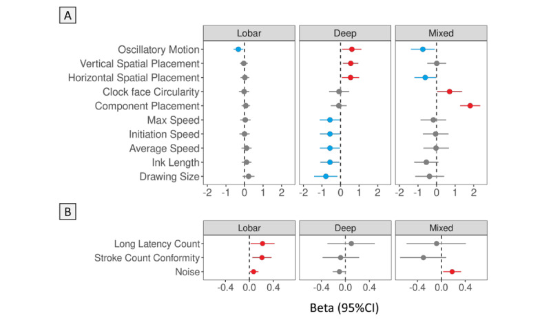
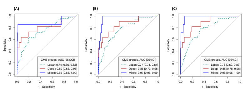
The study sample consisted of 1020 (n=585, 57.35% female) individuals aged 45 years and older (mean 72, SD 7.9 years). Among them, 64 (6.27%) participants exhibited CMB, comprising 46 with lobar-only, 11 with deep-only, and 7 with mixed (lobar+deep) CMB. Individuals with CMB tended to be older and had a higher prevalence of mild cognitive impairment and higher white matter hyperintensities compared to those without CMB (P<.05). While CMB were not associated with the paper-based clock-drawing test, participants with CMB had a lower overall DCT score (CMB: mean 68, SD 23 vs non-CMB: mean 76, SD 20; P=.009) in the univariate comparison. In the robust multiple regression model adjusted for covariates, deep CMB were significantly associated with lower scores on the drawing efficiency (β=-0.65, 95% CI -1.15 to -0.15; P=.01) and simple motor (β=-0.86, 95% CI -1.43 to -0.30; P=.003) domains of the command DCT. In the ROC curve analysis, DCT facets discriminated between no CMB and the CMB subtypes. The area under the ROC curve was 0.76 (95% CI 0.69-0.83) for lobar CMB, 0.88 (95% CI 0.78-0.98) for deep CMB, and 0.98 (95% CI 0.96-1.00) for mixed CMB, where the area under the ROC curve value nearing 1 indicated an accurate model.
The study indicates a significant association between CMB, especially deep and mixed types, and reduced performance in drawing efficiency and motor skills as assessed by the DCT. This highlights the potential of the DCT for early detection of CMB and their subtypes, providing a reliable alternative for cognitive assessment and making it a valuable tool for primary care screening before neuroimaging referral.
脑微出血(CMB)会增加患阿尔茨海默病的风险。目前用于检测 CMB 的神经影像学方法既昂贵又并非总是可及。
本研究旨在探讨数字时钟绘制测试(DCT)是否可以提供 CMB 的行为指标。
本研究分析了接受脑部磁共振成像扫描(西门子 1.5T,西门子医疗私人有限公司;T2*-GRE 加权序列)以诊断 CMB 并进行 DCT 预测的弗雷明汉心脏研究后代队列参与者的数据。此外,还在 DCT 期间收集了纸质时钟绘制测试。排除有痴呆或中风病史的个体。使用稳健的多变量线性回归模型来检查 DCT 方面得分与 CMB 患病率之间的关联,调整了相关协变量。使用接收者操作特征(ROC)曲线分析来评估 DCT 方面得分作为 CMB 患病率的预测因子。通过进一步纳入有中风和痴呆的参与者进行敏感性分析。
研究样本包括 1020 名(n=585,57.35%为女性)年龄在 45 岁及以上的个体(平均 72 岁,标准差 7.9 岁)。其中,64 名(6.27%)参与者出现 CMB,包括 46 名仅有皮质下 CMB、11 名仅有深部 CMB 和 7 名混合(皮质下+深部)CMB。与无 CMB 的参与者相比,有 CMB 的参与者年龄更大,患有轻度认知障碍的比例更高,且脑白质高信号更严重(P<.05)。虽然 CMB 与纸质时钟绘制测试无关联,但与无 CMB 的参与者相比,有 CMB 的参与者的总体 DCT 评分较低(CMB:平均 68,标准差 23 vs 无 CMB:平均 76,标准差 20;P=.009)。在调整协变量的稳健多元回归模型中,深部 CMB 与绘图效率(β=-0.65,95%CI -1.15 至 -0.15;P=.01)和简单运动(β=-0.86,95%CI -1.43 至 -0.30;P=.003)领域的指令 DCT 得分较低显著相关。在 ROC 曲线分析中,DCT 方面区分了无 CMB 和 CMB 亚型。ROC 曲线下面积为皮质下 CMB 为 0.76(95%CI 0.69-0.83),深部 CMB 为 0.88(95%CI 0.78-0.98),混合 CMB 为 0.98(95%CI 0.96-1.00),ROC 曲线下面积值接近 1 表示模型准确。
该研究表明 CMB(尤其是深部和混合性 CMB)与绘图效率和运动技能降低之间存在显著关联,这可通过 DCT 进行评估。这突出了 DCT 用于早期检测 CMB 及其亚型的潜力,为认知评估提供了可靠的替代方法,并使其成为神经影像学转诊前初级保健筛查的有价值工具。