• 文献检索
  • 文档翻译
  • 深度研究
  • 学术资讯
  • Suppr Zotero 插件Zotero 插件
  • 邀请有礼
  • 套餐&价格
  • 历史记录
应用&插件
Suppr Zotero 插件Zotero 插件浏览器插件Mac 客户端Windows 客户端微信小程序
定价
高级版会员购买积分包购买API积分包
服务
文献检索文档翻译深度研究API 文档MCP 服务
关于我们
关于 Suppr公司介绍联系我们用户协议隐私条款
关注我们

Suppr 超能文献

核心技术专利:CN118964589B侵权必究
粤ICP备2023148730 号-1Suppr @ 2026

文献检索

告别复杂PubMed语法,用中文像聊天一样搜索,搜遍4000万医学文献。AI智能推荐,让科研检索更轻松。

立即免费搜索

文件翻译

保留排版,准确专业,支持PDF/Word/PPT等文件格式,支持 12+语言互译。

免费翻译文档

深度研究

AI帮你快速写综述,25分钟生成高质量综述,智能提取关键信息,辅助科研写作。

立即免费体验

简单模型与深度学习在通过心电图检测低射血分数方面的比较。

Simple models vs. deep learning in detecting low ejection fraction from the electrocardiogram.

作者信息

Hughes John Weston, Somani Sulaiman, Elias Pierre, Tooley James, Rogers Albert J, Poterucha Timothy, Haggerty Christopher M, Salerno Michael, Ouyang David, Ashley Euan, Zou James, Perez Marco V

机构信息

Department of Computer Science, Stanford University, 353 Jane Stanford Way, Stanford, CA 94305, USA.

Department of Medicine, Stanford University, 1265 Pasteur Dr, Stanford, CA 94305, USA.

出版信息

Eur Heart J Digit Health. 2024 Apr 25;5(4):427-434. doi: 10.1093/ehjdh/ztae034. eCollection 2024 Jul.

DOI:10.1093/ehjdh/ztae034
PMID:39081946
原文链接:https://pmc.ncbi.nlm.nih.gov/articles/PMC11284011/
Abstract

AIMS

Deep learning methods have recently gained success in detecting left ventricular systolic dysfunction (LVSD) from electrocardiogram (ECG) waveforms. Despite their high level of accuracy, they are difficult to interpret and deploy broadly in the clinical setting. In this study, we set out to determine whether simpler models based on standard ECG measurements could detect LVSD with similar accuracy to that of deep learning models.

METHODS AND RESULTS

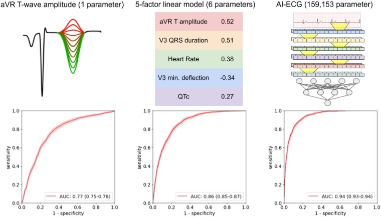
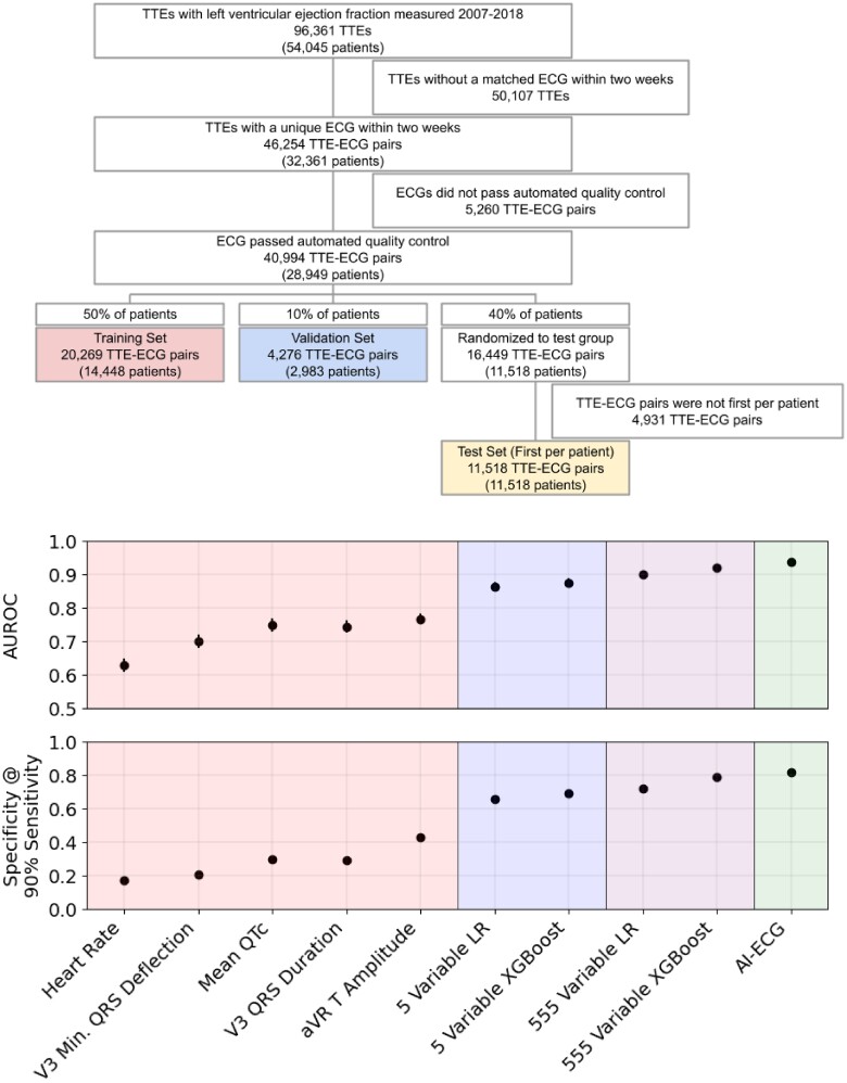
Using an observational data set of 40 994 matched 12-lead ECGs and transthoracic echocardiograms, we trained a range of models with increasing complexity to detect LVSD based on ECG waveforms and derived measurements. The training data were acquired from the Stanford University Medical Center. External validation data were acquired from the Columbia Medical Center and the UK Biobank. The Stanford data set consisted of 40 994 matched ECGs and echocardiograms, of which 9.72% had LVSD. A random forest model using 555 discrete, automated measurements achieved an area under the receiver operator characteristic curve (AUC) of 0.92 (0.91-0.93), similar to a deep learning waveform model with an AUC of 0.94 (0.93-0.94). A logistic regression model based on five measurements achieved high performance [AUC of 0.86 (0.85-0.87)], close to a deep learning model and better than N-terminal prohormone brain natriuretic peptide (NT-proBNP). Finally, we found that simpler models were more portable across sites, with experiments at two independent, external sites.

CONCLUSION

Our study demonstrates the value of simple electrocardiographic models that perform nearly as well as deep learning models, while being much easier to implement and interpret.

摘要

目的

深度学习方法最近在从心电图(ECG)波形中检测左心室收缩功能障碍(LVSD)方面取得了成功。尽管其准确性很高,但它们难以解释且难以在临床环境中广泛应用。在本研究中,我们着手确定基于标准ECG测量的更简单模型是否能以与深度学习模型相似的准确性检测LVSD。

方法和结果

使用包含40994对匹配的12导联ECG和经胸超声心动图的观察数据集,我们训练了一系列复杂度不断增加的模型,以基于ECG波形和派生测量来检测LVSD。训练数据来自斯坦福大学医学中心。外部验证数据来自哥伦比亚医学中心和英国生物银行。斯坦福数据集由40994对匹配的ECG和超声心动图组成,其中9.72%有LVSD。使用555个离散自动测量值的随机森林模型在受试者工作特征曲线(AUC)下的面积为0.92(0.91 - 0.93),与AUC为0.94(0.93 - 0.94)的深度学习波形模型相似。基于五项测量的逻辑回归模型具有高性能[AUC为0.86(0.85 - 0.87)],接近深度学习模型且优于N末端脑钠肽前体(NT - proBNP)。最后,我们发现更简单的模型在不同地点之间更具可移植性,并在两个独立的外部地点进行了实验。

结论

我们的研究证明了简单心电图模型的价值,其性能与深度学习模型相近,同时更易于实施和解释。

https://cdn.ncbi.nlm.nih.gov/pmc/blobs/f774/11284011/3ecc3f0c9656/ztae034f2.jpg
https://cdn.ncbi.nlm.nih.gov/pmc/blobs/f774/11284011/414b0aa75e9e/ztae034_ga.jpg
https://cdn.ncbi.nlm.nih.gov/pmc/blobs/f774/11284011/414b0aa75e9e/ztae034f1.jpg
https://cdn.ncbi.nlm.nih.gov/pmc/blobs/f774/11284011/3ecc3f0c9656/ztae034f2.jpg
https://cdn.ncbi.nlm.nih.gov/pmc/blobs/f774/11284011/414b0aa75e9e/ztae034_ga.jpg
https://cdn.ncbi.nlm.nih.gov/pmc/blobs/f774/11284011/414b0aa75e9e/ztae034f1.jpg
https://cdn.ncbi.nlm.nih.gov/pmc/blobs/f774/11284011/3ecc3f0c9656/ztae034f2.jpg

相似文献

1
Simple models vs. deep learning in detecting low ejection fraction from the electrocardiogram.简单模型与深度学习在通过心电图检测低射血分数方面的比较。
Eur Heart J Digit Health. 2024 Apr 25;5(4):427-434. doi: 10.1093/ehjdh/ztae034. eCollection 2024 Jul.
2
External validation of a deep learning electrocardiogram algorithm to detect ventricular dysfunction.深度学习心电图算法检测心室功能障碍的外部验证。
Int J Cardiol. 2021 Apr 15;329:130-135. doi: 10.1016/j.ijcard.2020.12.065. Epub 2021 Jan 2.
3
Deep learning algorithm for predicting left ventricular systolic dysfunction in atrial fibrillation with rapid ventricular response.用于预测快速心室反应型心房颤动患者左心室收缩功能障碍的深度学习算法
Eur Heart J Digit Health. 2024 Aug 19;5(6):683-691. doi: 10.1093/ehjdh/ztae062. eCollection 2024 Nov.
4
Artificial intelligence-enabled electrocardiographic screening for left ventricular systolic dysfunction and mortality risk prediction.基于人工智能的心电图筛查左心室收缩功能障碍及死亡风险预测
Front Cardiovasc Med. 2023 Mar 3;10:1070641. doi: 10.3389/fcvm.2023.1070641. eCollection 2023.
5
Artificial Intelligence-Enabled ECG Algorithm to Identify Patients With Left Ventricular Systolic Dysfunction Presenting to the Emergency Department With Dyspnea.基于人工智能的心电图算法识别因呼吸困难就诊于急诊科的左心室收缩功能障碍患者。
Circ Arrhythm Electrophysiol. 2020 Aug;13(8):e008437. doi: 10.1161/CIRCEP.120.008437. Epub 2020 Aug 4.
6
Ensemble Deep Learning Algorithm for Structural Heart Disease Screening Using Electrocardiographic Images: PRESENT SHD.用于使用心电图图像进行结构性心脏病筛查的集成深度学习算法:呈现结构性心脏病。
J Am Coll Cardiol. 2025 Apr 1;85(12):1302-1313. doi: 10.1016/j.jacc.2025.01.030.
7
Development and validation of an electrocardiographic artificial intelligence model for detection of peripartum cardiomyopathy.开发和验证一种心电图人工智能模型,用于检测围产期心肌病。
Am J Obstet Gynecol MFM. 2024 Apr;6(4):101337. doi: 10.1016/j.ajogmf.2024.101337. Epub 2024 Mar 4.
8
Artificial intelligence-based identification of left ventricular systolic dysfunction from 12-lead electrocardiograms: external validation and advanced application of an existing model.基于人工智能从12导联心电图识别左心室收缩功能障碍:现有模型的外部验证及进阶应用
Eur Heart J Digit Health. 2023 Dec 20;5(2):144-151. doi: 10.1093/ehjdh/ztad081. eCollection 2024 Mar.
9
Left ventricular systolic dysfunction predicted by artificial intelligence using the electrocardiogram in Chagas disease patients-The SaMi-Trop cohort.人工智能利用心电图预测恰加斯病患者的左心室收缩功能障碍——SaMi-Trop 队列研究。
PLoS Negl Trop Dis. 2021 Dec 6;15(12):e0009974. doi: 10.1371/journal.pntd.0009974. eCollection 2021 Dec.
10
Artificial intelligence-enabled electrocardiogram screens low left ventricular ejection fraction with a degree of confidence.人工智能驱动的心电图能够以一定的置信度筛查出左心室射血分数较低的情况。
Digit Health. 2022 Dec 12;8:20552076221143249. doi: 10.1177/20552076221143249. eCollection 2022 Jan-Dec.

引用本文的文献

1
A deep learning phenome wide association study of the electrocardiogram.一项关于心电图的深度学习全表型关联研究。
Eur Heart J Digit Health. 2025 May 8;6(4):595-607. doi: 10.1093/ehjdh/ztaf047. eCollection 2025 Jul.
2
AI analysis for ejection fraction estimation from 12-lead ECG.基于12导联心电图估计射血分数的人工智能分析。
Sci Rep. 2025 Apr 18;15(1):13502. doi: 10.1038/s41598-025-97113-0.

本文引用的文献

1
Race, Sex, and Age Disparities in the Performance of ECG Deep Learning Models Predicting Heart Failure.种族、性别和年龄差异对心电图深度学习模型预测心力衰竭性能的影响。
Circ Heart Fail. 2024 Jan;17(1):e010879. doi: 10.1161/CIRCHEARTFAILURE.123.010879. Epub 2023 Dec 21.
2
Development and validation of a rapid visual technique for left ventricular hypertrophy detection from the electrocardiogram.一种用于从心电图检测左心室肥厚的快速视觉技术的开发与验证。
Front Cardiovasc Med. 2023 Aug 23;10:1251511. doi: 10.3389/fcvm.2023.1251511. eCollection 2023.
3
A deep learning-based electrocardiogram risk score for long term cardiovascular death and disease.
一种基于深度学习的长期心血管死亡和疾病的心电图风险评分
NPJ Digit Med. 2023 Sep 12;6(1):169. doi: 10.1038/s41746-023-00916-6.
4
Artificial Intelligence and Machine Learning in Clinical Medicine, 2023.临床医学中的人工智能与机器学习,2023年。
N Engl J Med. 2023 Mar 30;388(13):1201-1208. doi: 10.1056/NEJMra2302038.
5
Deep Learning Electrocardiographic Analysis for Detection of Left-Sided Valvular Heart Disease.深度学习心电图分析用于左心瓣膜性心脏病的检测。
J Am Coll Cardiol. 2022 Aug 9;80(6):613-626. doi: 10.1016/j.jacc.2022.05.029.
6
Relation of T Wave Positivity in Lead aVR to Ischemic Etiology of Cardiomyopathy.aVR 导联 T 波正向与心肌病缺血性病因的关系。
Am J Cardiol. 2022 Oct 1;180:17-23. doi: 10.1016/j.amjcard.2022.06.043. Epub 2022 Jul 29.
7
Performance of a Convolutional Neural Network and Explainability Technique for 12-Lead Electrocardiogram Interpretation.卷积神经网络性能及 12 导联心电图解释的可解释性技术。
JAMA Cardiol. 2021 Nov 1;6(11):1285-1295. doi: 10.1001/jamacardio.2021.2746.
8
Electrocardiographic biomarkers to predict atrial fibrillation in sinus rhythm electrocardiograms.心电图生物标志物预测窦性心律心电图中的心房颤动。
Heart. 2021 Nov;107(22):1813-1819. doi: 10.1136/heartjnl-2021-319120. Epub 2021 Jun 4.
9
Prediction of mortality from 12-lead electrocardiogram voltage data using a deep neural network.利用深度神经网络预测 12 导联心电图电压数据的死亡率。
Nat Med. 2020 Jun;26(6):886-891. doi: 10.1038/s41591-020-0870-z. Epub 2020 May 11.
10
Key challenges for delivering clinical impact with artificial intelligence.人工智能实现临床影响的关键挑战。
BMC Med. 2019 Oct 29;17(1):195. doi: 10.1186/s12916-019-1426-2.