Mun Aung Ye, Akiyama Kentaro, Wang Ziyi, Zhang Jiewen, Kitagawa Wakana, Kohno Teisaku, Tagashira Ryuji, Ishibashi Kei, Matsunaga Naoya, Zou Tingling, Ono Mitsuaki, Kuboki Takuo
Department of Oral Rehabilitation and Regenerative Medicine, Graduate School of Medicine, Dentistry and Pharmaceutical Sciences, Okayama University, 2-5-1 Shikata-cho, Kita-ku, Okayama 700-8525, Japan.
Department of Molecular Biology and Biochemistry, Graduate School of Medicine, Dentistry and Pharmaceutical Sciences, Okayama University, 2-5-1 Shikata-cho, Kita-ku, Okayama 700-8558, Japan.
JBMR Plus. 2024 Jul 4;8(8):ziae085. doi: 10.1093/jbmrpl/ziae085. eCollection 2024 Aug.
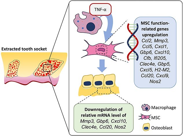
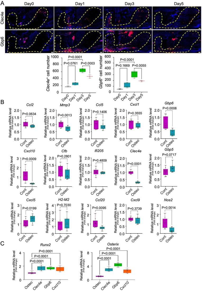
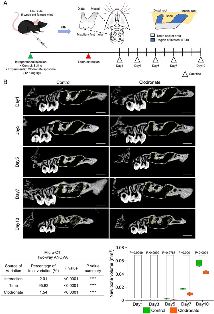
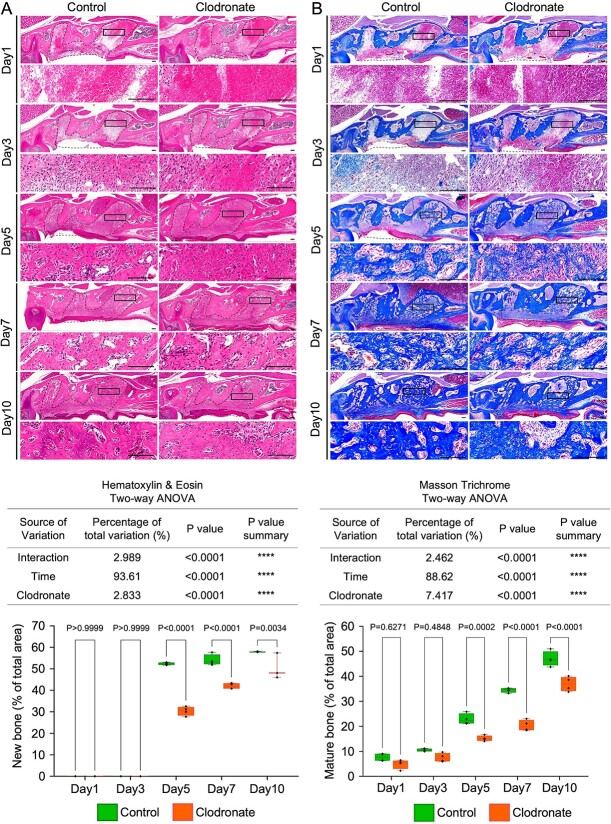
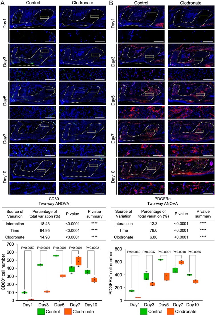
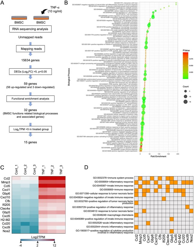
Mesenchymal stem cells (MSCs) and macrophages collaboratively contribute to bone regeneration after injury. However, detailed mechanisms underlying the interaction between MSCs and inflammatory macrophages (M1) remain unclear. A macrophage-depleted tooth extraction model was generated in 5-wk-old female C57BL/6J mice using clodronate liposome (12.5 mg/kg/mouse, intraperitoneally) or saline injection (control) before maxillary first molar extraction. Mice were sacrificed on days 1, 3, 5, 7, and 10 after tooth extraction ( = 4). Regenerated bone volume evaluation of tooth extraction socket (TES) and histochemical analysis of CD80M1, CD206M2 (anti-inflammatory macrophages), PDGFRαMSC, and TNF-α cells were performed. In vitro, isolated MSCs with or without TNF-α stimulation (10 ng/mL, 24 h, = 3) were bulk RNA-sequenced (RNA-Seq) to identify TNF-α stimulation-specific MSC transcriptomes. Day 7 micro-CT and HE staining revealed significantly lower mean bone volume (clodronate vs control: 0.01 mm vs 0.02 mm, <.0001) and mean percentage of regenerated bone area per total TES in clodronate group (41.97% vs 54.03%, <.0001). Clodronate group showed significant reduction in mean number of CD80, TNF-α, PDGFRα, and CD80TNF-α cells on day 5 (306.5 vs 558.8, <.0001; 280.5 vs 543.8, <.0001; 365.0 vs 633.0, <.0001, 29.0 vs 42.5, <.0001), while these cells recovered significantly on day 7 (493.3 vs 396.0, =.0004; 479.3 vs 384.5, =.0008; 593.0 vs 473.0, =.0010, 41.0 vs 32.5, =.0003). RNA-Seq analysis showed that 15 genes (|log2FC| > 5.0, log2TPM > 5) after TNF-α stimulation were candidates for regulating MSC's immunomodulatory capacity. In vivo, and are involved in inflammation and bone formation. , , and knockdown increased osteogenic differentiation of MSCs in vitro. Temporal reduction followed by apparent recovery of TNF-α-producing M1 macrophages and MSCs after temporal macrophage depletion suggests that TNF-α activated MSCs during TES healing. In vitro mimicking the effect of TNF-α on MSCs indicated that there are 15 candidate MSC genes for regulation of immunomodulatory capacity.
间充质干细胞(MSCs)和巨噬细胞共同促进损伤后的骨再生。然而,MSCs与炎性巨噬细胞(M1)之间相互作用的详细机制仍不清楚。在5周龄雌性C57BL/6J小鼠中,于拔除上颌第一磨牙前,使用氯膦酸盐脂质体(12.5mg/kg/小鼠,腹腔注射)或注射生理盐水(对照)建立巨噬细胞耗竭的拔牙模型。在拔牙后第1、3、5、7和10天处死小鼠(每组n = 4)。对拔牙窝(TES)进行再生骨体积评估,并对CD80(M1)、CD206(M2,抗炎巨噬细胞)、PDGFRα(MSCs)和TNF-α细胞进行组织化学分析。在体外,对有或无TNF-α刺激(10ng/mL,24小时,每组n = 3)的分离MSCs进行批量RNA测序(RNA-Seq),以鉴定TNF-α刺激特异性的MSCs转录组。第7天的显微CT和苏木精-伊红染色显示,氯膦酸盐组的平均骨体积显著降低(氯膦酸盐组与对照组:0.01mm对0.02mm,P <.0001),且氯膦酸盐组中再生骨面积占总TES的平均百分比也显著降低(41.97%对54.03%,P <.0001)。氯膦酸盐组在第5天时CD80、TNF-α、PDGFRα和CD80+TNF-α细胞的平均数量显著减少(306.5对558.8,P <.0001;280.5对543.8,P <.0001;365.0对633.0,P <.0001,29.0对42.5,P <.0001),而这些细胞在第7天时显著恢复(493.3对396.0,P = 0.0004;479.3对384.5,P = 0.0008;593.0对473.0,P = 0.0010,41.0对32.5,P = 0.0003)。RNA-Seq分析表明,TNF-α刺激后有15个基因(|log2FC| > 5.0,log2TPM > 5)是调节MSCs免疫调节能力的候选基因。在体内,[基因名称1]和[基因名称2]参与炎症和骨形成。[基因名称3]、[基因名称4]和[基因名称5]敲低可增加体外MSCs的成骨分化。巨噬细胞短暂耗竭后,产生TNF-α的M1巨噬细胞和MSCs先暂时减少随后明显恢复,这表明在TES愈合过程中TNF-α激活了MSCs。体外模拟TNF-α对MSCs的作用表明,有15个候选MSCs基因可调节免疫调节能力。