Bodden Jannis, Prucker Philipp, Sekuboyina Anjany, El Husseini Malek, Grau Katharina, Rühling Sebastian, Burian Egon, Zimmer Claus, Baum Thomas, Kirschke Jan S
Department of Neuroradiology, TUM School of Medicine, Klinikum rechts der Isar, Technical University of Munich, Munich, Germany.
Department of Informatics, TUM School of Computation, Information and Technology, Technical University of Munich, Munich, Germany.
Eur Radiol Exp. 2024 Aug 1;8(1):86. doi: 10.1186/s41747-024-00483-9.
To investigate the reproducibility of automated volumetric bone mineral density (vBMD) measurements from routine thoracoabdominal computed tomography (CT) assessed with segmentations by a convolutional neural network and automated correction of contrast phases, on diverse scanners, with scanner-specific asynchronous or scanner-agnostic calibrations.
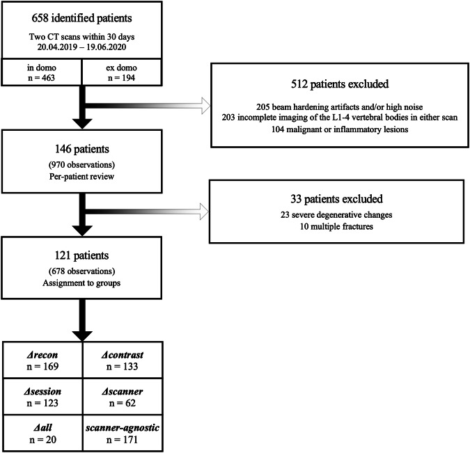
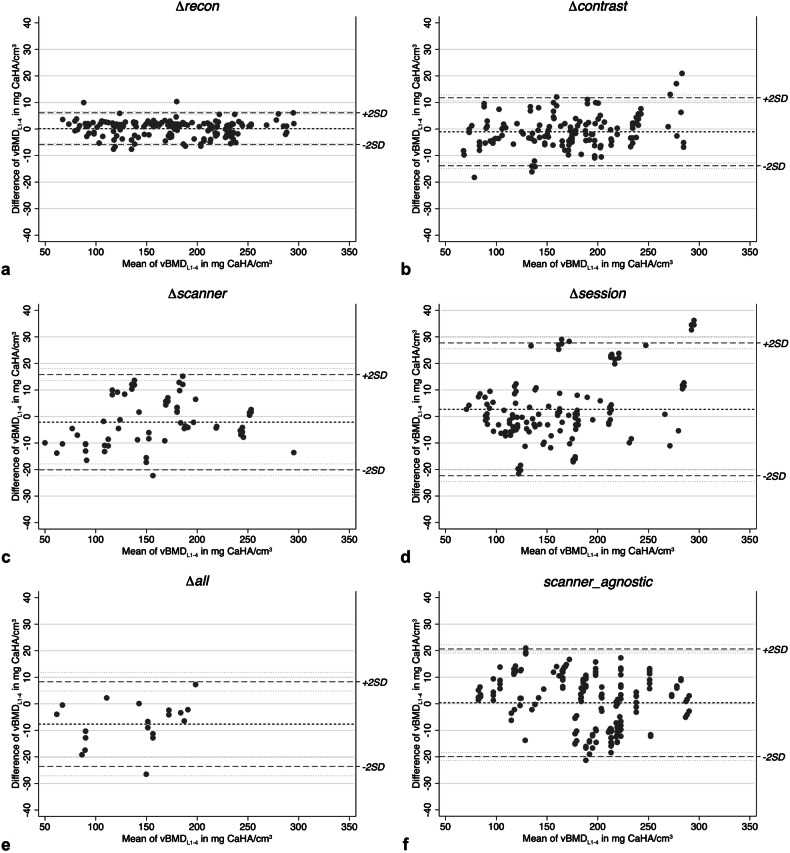
We obtained 679 observations from 278 CT scans in 121 patients (77 males, 63.6%) studied from 04/2019 to 06/2020. Observations consisted of two vBMD measurements from Δdifferent reconstruction kernels (n = 169), Δcontrast phases (n = 133), scan Δsessions (n = 123), Δscanners (n = 63), or Δall of the aforementioned (n = 20), and observations lacking scanner-specific calibration (n = 171). Precision was assessed using root-mean-square error (RMSE) and root-mean-square coefficient of variation (RMSCV). Cross-measurement agreement was assessed using Bland-Altman plots; outliers within 95% confidence interval of the limits of agreement were reviewed.
Repeated measurements from Δdifferent reconstruction kernels were highly precise (RMSE 3.0 mg/cm; RMSCV 1.3%), even for consecutive scans with different Δcontrast phases (RMSCV 2.9%). Measurements from different Δscan sessions or Δscanners showed decreased precision (RMSCV 4.7% and 4.9%, respectively). Plot-review identified 12 outliers from different scan Δsessions, with signs of hydropic decompensation. Observations with Δall differences showed decreased precision compared to those lacking scanner-specific calibration (RMSCV 5.9 and 3.7, respectively).
Automatic vBMD assessment from routine CT is precise across varying setups, when calibrated appropriately. Low precision was found in patients with signs of new or worsening hydropic decompensation, what should be considered an exclusion criterion for both opportunistic and dedicated quantitative CT.
Automated CT-based vBMD measurements are precise in various scenarios, including cross-session and cross-scanner settings, and may therefore facilitate opportunistic screening for osteoporosis and surveillance of BMD in patients undergoing routine clinical CT scans.
Artificial intelligence-based tools facilitate BMD measurements in routine clinical CT datasets. Automated BMD measurements are highly reproducible in various settings. Reliable, automated opportunistic osteoporosis diagnostics allow for large-scale application.
本研究旨在探讨通过卷积神经网络分割及造影剂相自动校正,利用常规胸腹计算机断层扫描(CT)进行自动容积骨密度(vBMD)测量的可重复性,涉及多种扫描仪,采用特定扫描仪的异步校准或与扫描仪无关的校准方法。
我们收集了2019年4月至2020年6月期间121例患者(77例男性,占63.6%)的278次CT扫描数据,共679份观察数据。观察数据包括来自不同重建核(n = 169)、不同造影剂相(n = 133)、不同扫描时段(n = 123)、不同扫描仪(n = 63)或上述所有因素均不同(n = 20)的两次vBMD测量数据,以及缺乏特定扫描仪校准的观察数据(n = 171)。使用均方根误差(RMSE)和均方根变异系数(RMSCV)评估精度。使用布兰德-奥特曼图评估交叉测量一致性;对一致性界限95%置信区间内的异常值进行审查。
来自不同重建核的重复测量具有高度精确性(RMSE 3.0mg/cm;RMSCV 1.3%),即使是不同造影剂相的连续扫描(RMSCV 2.9%)。来自不同扫描时段或不同扫描仪的测量精度有所下降(RMSCV分别为4.7%和4.9%)。通过图审查发现来自不同扫描时段的12个异常值,有水肿性失代偿的迹象。与缺乏特定扫描仪校准的观察数据相比,所有差异均存在的观察数据精度下降(RMSCV分别为5.9和3.7)。
在适当校准的情况下,常规CT自动vBMD评估在不同设置下是精确的。在有新发或加重的水肿性失代偿迹象的患者中发现精度较低,这应被视为机会性和专用定量CT的排除标准。
基于CT的自动vBMD测量在各种情况下都是精确的,包括跨时段和跨扫描仪设置,因此可能有助于在接受常规临床CT扫描的患者中进行机会性骨质疏松筛查和骨密度监测。
基于人工智能的工具有助于在常规临床CT数据集中进行骨密度测量。自动骨密度测量在各种设置下具有高度可重复性。可靠的自动机会性骨质疏松诊断允许大规模应用。