Department of Nephrology, The First Hospital of Jilin University, Changchun, China.
Department of Hematology, The First Hospital of Jilin University, Changchun, China.
Sci Rep. 2024 Aug 3;14(1):18010. doi: 10.1038/s41598-024-69104-0.
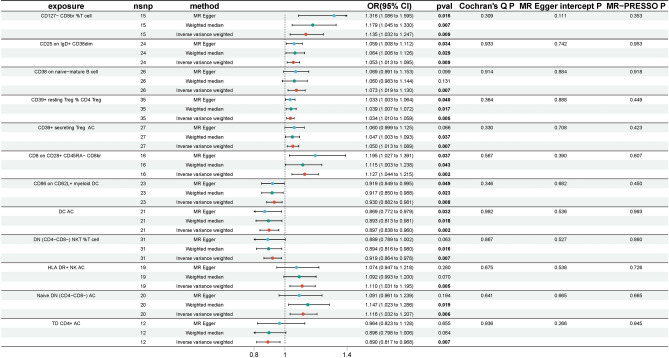
Prior research has identified associations between immune cells and aplastic anaemia (AA); however, the causal relationships between them have not been conclusively established. A two-sample Mendelian randomisation analysis was conducted to investigate the causal link between 731 immune cell signatures and AA risk using publicly available genetic data. Four types of immune signatures, including relative cell, absolute cell (AC), median fluorescence intensities and morphological parameters, were considered sensitivity analyses were also performed to verify the robustness of the results and assess potential issues such as heterogeneity and horizontal pleiotropy. Following multiple test adjustments using the False Discovery Rate (FDR) method, no statistically significant impact of any immunophenotype on AA was observed. However, twelve immunophenotypes exhibited a significant correlation with AA without FDR correction (p of IVW < 0.01), of which eight were harmful to AA: CD127- CD8br %T cell (Treg panel), CD25 on IgD + CD38dim (B cell panel), CD38 on naive-mature B cell (B cell panel), CD39 + resting Treg % CD4 Treg (Treg panel), CD39 + secreting Treg AC (Treg panel), CD8 on CD28 + CD45RA- CD8br (Treg panel), HLA DR + NK AC (TBNK panel), Naive DN (CD4CD8) AC (Maturation stages of T cell panel); and four were protective to AA: CD86 on CD62L + myeloid DC (cDC panel), DC AC (cDC panel), DN (CD4CD8) NKT %T cell (TBNK panel), and TD CD4 + AC (Maturation stages of T cell panel). The results of this study demonstrate a close link between immune cells and AA by genetic means, thereby improving the current understanding of the interaction between immune cells and AA risk and providing guidance for future clinical research.
先前的研究已经确定了免疫细胞与再生障碍性贫血(AA)之间的关联;然而,它们之间的因果关系尚未得到明确确立。本研究采用两样本孟德尔随机化分析,利用公开的遗传数据,研究了 731 种免疫细胞特征与 AA 风险之间的因果关系。考虑了四种类型的免疫特征,包括相对细胞、绝对细胞(AC)、中荧光强度和形态参数。还进行了敏感性分析,以验证结果的稳健性,并评估潜在问题,如异质性和水平多效性。使用 False Discovery Rate(FDR)方法进行多次测试调整后,没有观察到任何免疫表型对 AA 有统计学显著影响。然而,有 12 种免疫表型在未经 FDR 校正的情况下与 AA 显著相关(IVW 的 p 值<0.01),其中 8 种对 AA 有害:CD127-CD8br%T 细胞(Treg 面板)、IgD+CD38dim 上的 CD25(B 细胞面板)、幼稚-成熟 B 细胞上的 CD38(B 细胞面板)、CD39+静息 Treg%CD4 Treg(Treg 面板)、CD39+分泌 Treg AC(Treg 面板)、CD8 上的 CD28+CD45RA-CD8br(Treg 面板)、HLA DR+NK AC(TBNK 面板)、幼稚 DN(CD4CD8)AC(T 细胞成熟阶段面板);四种对 AA 有保护作用:CD62L+髓样 DC 上的 CD86(cDC 面板)、DC AC(cDC 面板)、DN(CD4CD8)NKT%T 细胞(TBNK 面板)和 TD CD4+AC(T 细胞成熟阶段面板)。本研究通过遗传手段证明了免疫细胞与 AA 之间的密切联系,从而提高了对免疫细胞与 AA 风险相互作用的现有认识,并为未来的临床研究提供了指导。