Department of Surgery, Mayo Clinic, Rochester, MN, United States.
Boston Children's Hospital, Harvard Medical School, Boston, MA, United States.
Front Immunol. 2024 Jul 22;15:1448092. doi: 10.3389/fimmu.2024.1448092. eCollection 2024.
The immunomodulatory properties of mesenchymal stromal cells (MSC) have been well-characterized in and models. We have previously shown that liver MSC (L-MSC) are superior inhibitors of T-cell activation/proliferation, NK cell cytolytic function, and macrophage activation compared to adipose (A-MSC) and bone marrow MSC (BM-MSC) .
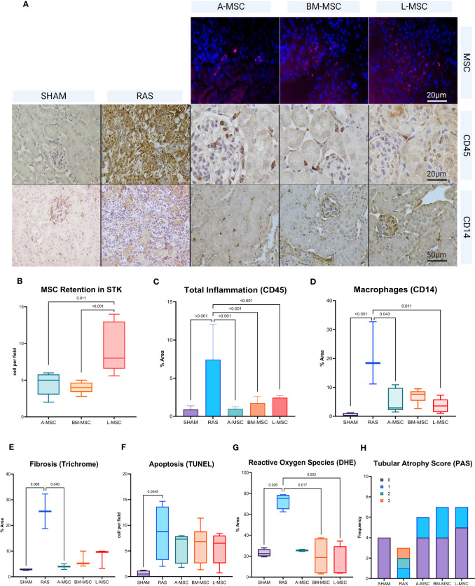
To test these observations , we infused these types of MSC into mice with unilateral renal artery stenosis (RAS), an established model of kidney inflammation. Unilateral RAS was induced via laparotomy in 11-week-old, male 129-S1 mice under general anesthesia. Control mice had sham operations. Human L-MSC, AMSC, and BM-MSC (5x10⁵ cells each) or PBS vehicle were injected intra-arterially 2 weeks after surgery. Kidney morphology was studied 2 weeks after infusion using micro-MRI imaging. Renal inflammation, apoptosis, fibrosis, and MSC retention were studied utilizing western blot, immunofluorescence, and immunohistological analyses.
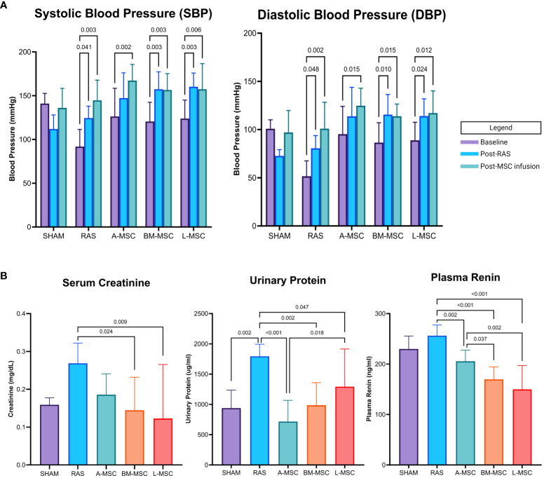
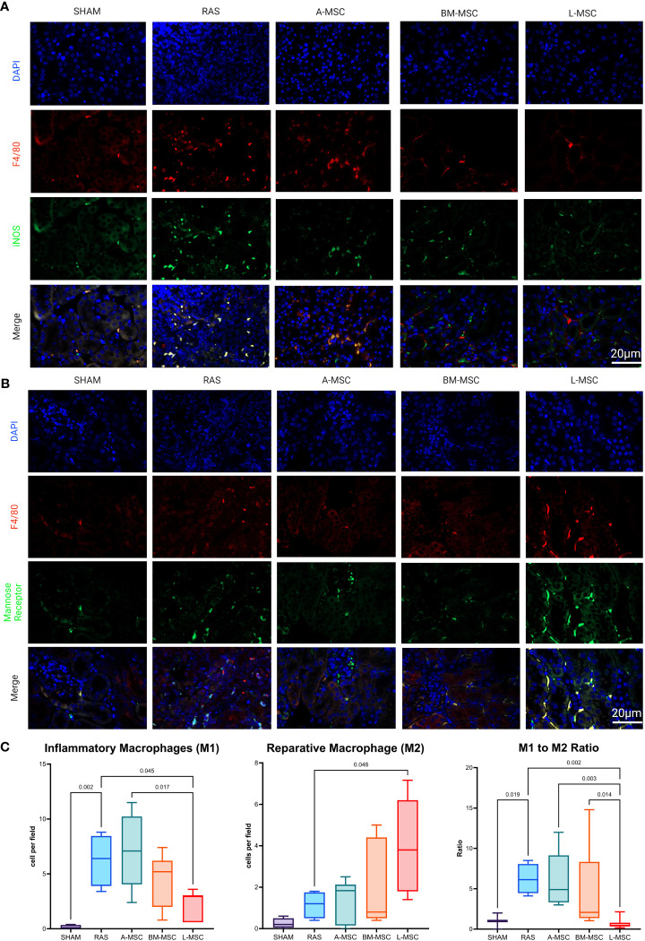
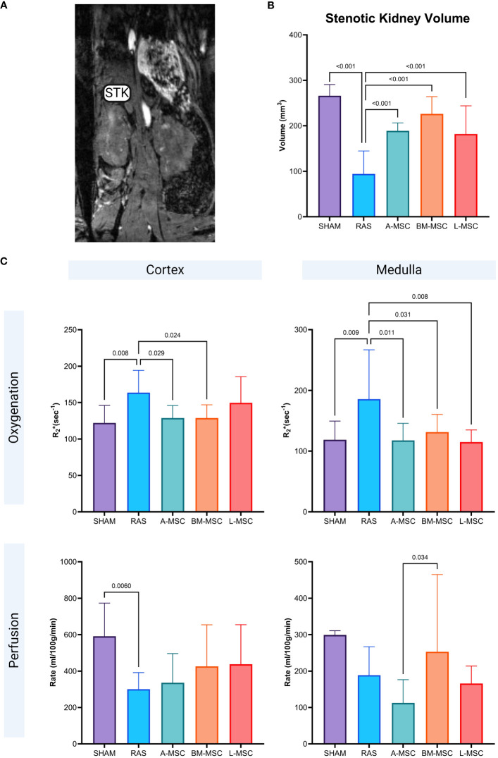
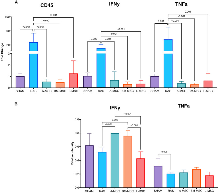
The stenotic kidney volume was smaller in all RAS mice, confirming significant injury, and was improved by infusion of all MSC types. All MSC-infused groups had lower levels of plasma renin and proteinuria compared to untreated RAS. Serum creatinine improved in micetreated with BM- and L-MSC. All types of MSC located to and were retained within the stenotic kidneys, but L-MSC retention was significantly higher than A- and BM-MSC. While all groups of MSC-treated mice displayed reduced overall inflammation and macrophage counts, L-MSC showed superior potency at localizing to the site of inflammation and inducing M2 (reparative) macrophage polarization to reduce inflammatory changes.
These findings extend our studies and suggest that L-MSC possess unique anti-inflammatory properties that may play a role in liver-induced tolerance and lend further support to their use as therapeutic agents for diseases with underlying inflammatory pathophysiology.
间充质基质细胞(MSC)的免疫调节特性在 和 模型中得到了很好的描述。我们之前已经表明,与脂肪(A-MSC)和骨髓 MSC(BM-MSC)相比,肝 MSC(L-MSC)是 T 细胞激活/增殖、NK 细胞细胞溶解功能和巨噬细胞激活的更好抑制剂 。
为了验证这些观察结果,我们将这些类型的 MSC 输注到单侧肾动脉狭窄(RAS)的小鼠中,这是一种已建立的肾脏炎症模型。在全身麻醉下,通过剖腹手术在 11 周龄雄性 129-S1 小鼠中诱导单侧 RAS。对照小鼠接受假手术。在手术后 2 周,通过动脉内注射 5x10⁵ 个每个细胞的人 L-MSC、A-MSC 和 BM-MSC 或 PBS 载体。在输注后 2 周,使用 micro-MRI 成像研究肾脏形态。利用 Western blot、免疫荧光和免疫组织化学分析研究肾炎症、细胞凋亡、纤维化和 MSC 保留。
所有 RAS 小鼠的狭窄肾脏体积均较小,证实了严重损伤,并通过输注所有 MSC 类型得到改善。与未治疗的 RAS 相比,所有 MSC 输注组的血浆肾素和蛋白尿水平均较低。BM-和 L-MSC 治疗的小鼠血清肌酐水平改善。所有类型的 MSC 均定位于狭窄肾脏并保留在其中,但 L-MSC 的保留率明显高于 A-MSC 和 BM-MSC。虽然所有 MSC 治疗组的小鼠均表现出总体炎症和巨噬细胞计数减少,但 L-MSC 显示出优越的定位到炎症部位和诱导 M2(修复)巨噬细胞极化以减少炎症变化的能力。
这些发现扩展了我们的研究,并表明 L-MSC 具有独特的抗炎特性,可能在肝诱导的耐受中发挥作用,并进一步支持将其用作潜在炎症发病机制疾病的治疗剂。