• 文献检索
  • 文档翻译
  • 深度研究
  • 学术资讯
  • Suppr Zotero 插件Zotero 插件
  • 邀请有礼
  • 套餐&价格
  • 历史记录
应用&插件
Suppr Zotero 插件Zotero 插件浏览器插件Mac 客户端Windows 客户端微信小程序
定价
高级版会员购买积分包购买API积分包
服务
文献检索文档翻译深度研究API 文档MCP 服务
关于我们
关于 Suppr公司介绍联系我们用户协议隐私条款
关注我们

Suppr 超能文献

核心技术专利:CN118964589B侵权必究
粤ICP备2023148730 号-1Suppr @ 2026

文献检索

告别复杂PubMed语法,用中文像聊天一样搜索,搜遍4000万医学文献。AI智能推荐,让科研检索更轻松。

立即免费搜索

文件翻译

保留排版,准确专业,支持PDF/Word/PPT等文件格式,支持 12+语言互译。

免费翻译文档

深度研究

AI帮你快速写综述,25分钟生成高质量综述,智能提取关键信息,辅助科研写作。

立即免费体验

通过视神经乳头和黄斑光学相干断层扫描厚度测量检测视野恶化

Detecting Visual Field Worsening From Optic Nerve Head and Macular Optical Coherence Tomography Thickness Measurements.

作者信息

Pham Alex T, Pan Annabelle A, Bradley Chris, Hou Kaihua, Herbert Patrick, Johnson Chris, Wall Michael, Yohannan Jithin

机构信息

Wilmer Eye Institute, Johns Hopkins University School of Medicine, Baltimore, MD, USA.

Malone Center for Engineering in Healthcare, Johns Hopkins University, Baltimore, MD, USA.

出版信息

Transl Vis Sci Technol. 2024 Aug 1;13(8):12. doi: 10.1167/tvst.13.8.12.

DOI:10.1167/tvst.13.8.12
PMID:39115839
原文链接:https://pmc.ncbi.nlm.nih.gov/articles/PMC11316451/
Abstract

PURPOSE

Compare the use of optic disc and macular optical coherence tomography measurements to predict glaucomatous visual field (VF) worsening.

METHODS

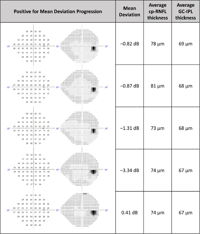
Machine learning and statistical models were trained on 924 eyes (924 patients) with circumpapillary retinal nerve fiber layer (cp-RNFL) or ganglion cell inner plexiform layer (GC-IPL) thickness measurements. The probability of 24-2 VF worsening was predicted using both trend-based and event-based progression definitions of VF worsening. Additionally, the cp-RNFL and GC-IPL predictions were combined to produce a combined prediction. A held-out test set of 617 eyes was used to calculate the area under the curve (AUC) to compare cp-RNFL, GC-IPL, and combined predictions.

RESULTS

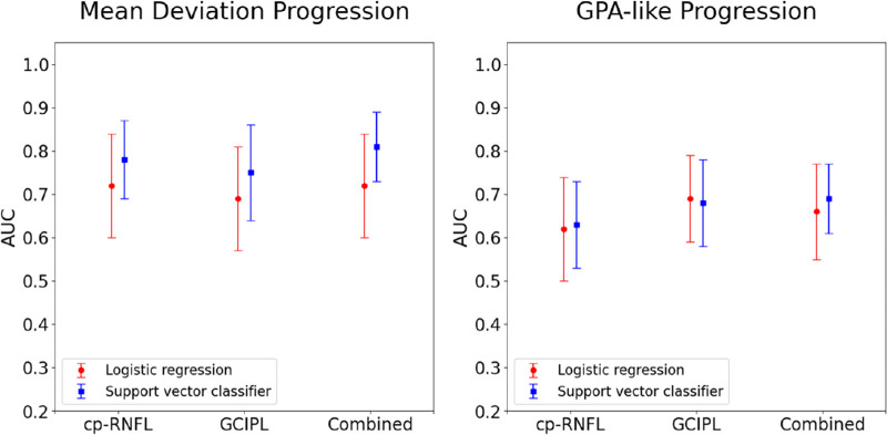
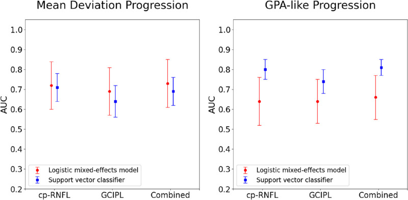
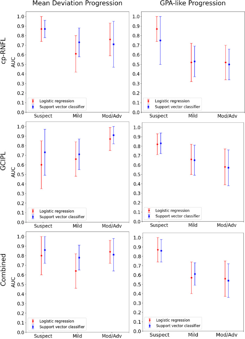
The AUCs for cp-RNFL, GC-IPL, and combined predictions with the statistical and machine learning models were 0.72, 0.69, 0.73, and 0.78, 0.75, 0.81, respectively, when using trend-based analysis as ground truth. The differences in performance between the cp-RNFL, GC-IPL, and combined predictions were not statistically significant. AUCs were highest in glaucoma suspects using cp-RNFL predictions and highest in moderate/advanced glaucoma using GC-IPL predictions. The AUCs for the statistical and machine learning models were 0.63, 0.68, 0.69, and 0.72, 0.69, 0.73, respectively, when using event-based analysis. AUCs decreased with increasing disease severity for all predictions.

CONCLUSIONS

cp-RNFL and GC-IPL similarly predicted VF worsening overall, but cp-RNFL performed best in early glaucoma stages and GC-IPL in later stages. Combining both did not enhance detection significantly.

TRANSLATIONAL RELEVANCE

cp-RNFL best predicted trend-based 24-2 VF progression in early-stage disease, while GC-IPL best predicted progression in late-stage disease. Combining both features led to minimal improvement in predicting progression.

摘要

目的

比较视盘和黄斑光学相干断层扫描测量用于预测青光眼性视野(VF)恶化的情况。

方法

对924只眼(924例患者)进行机器学习和统计模型训练,这些患者有视乳头周围视网膜神经纤维层(cp-RNFL)或神经节细胞内丛状层(GC-IPL)厚度测量值。使用基于趋势和基于事件的VF恶化进展定义来预测24-2 VF恶化的概率。此外,将cp-RNFL和GC-IPL预测结果相结合以产生联合预测。使用617只眼的保留测试集来计算曲线下面积(AUC),以比较cp-RNFL、GC-IPL和联合预测结果。

结果

当使用基于趋势的分析作为金标准时,cp-RNFL、GC-IPL以及联合统计模型和机器学习模型的预测的AUC分别为0.72、0.69、0.73以及0.78、0.75、0.81。cp-RNFL、GC-IPL和联合预测之间的性能差异无统计学意义。在青光眼可疑患者中,使用cp-RNFL预测时AUC最高,在中度/重度青光眼患者中,使用GC-IPL预测时AUC最高。当使用基于事件的分析时,统计模型和机器学习模型的AUC分别为0.63、0.68、0.69以及0.72、0.69、0.73。所有预测的AUC均随疾病严重程度增加而降低。

结论

cp-RNFL和GC-IPL总体上对VF恶化的预测相似,但cp-RNFL在青光眼早期阶段表现最佳,而GC-IPL在后期阶段表现最佳。两者结合并不能显著提高检测效果。

转化相关性

cp-RNFL最能预测早期疾病中基于趋势的24-2 VF进展,而GC-IPL最能预测晚期疾病的进展。结合这两个特征在预测进展方面的改善最小。

https://cdn.ncbi.nlm.nih.gov/pmc/blobs/41ca/11316451/1ae40e42bc43/tvst-13-8-12-f005.jpg
https://cdn.ncbi.nlm.nih.gov/pmc/blobs/41ca/11316451/45d408f18232/tvst-13-8-12-f001.jpg
https://cdn.ncbi.nlm.nih.gov/pmc/blobs/41ca/11316451/ec3145042c7b/tvst-13-8-12-f002.jpg
https://cdn.ncbi.nlm.nih.gov/pmc/blobs/41ca/11316451/c79ccb003204/tvst-13-8-12-f003.jpg
https://cdn.ncbi.nlm.nih.gov/pmc/blobs/41ca/11316451/4834df1ea237/tvst-13-8-12-f004.jpg
https://cdn.ncbi.nlm.nih.gov/pmc/blobs/41ca/11316451/1ae40e42bc43/tvst-13-8-12-f005.jpg

相似文献

1
Detecting Visual Field Worsening From Optic Nerve Head and Macular Optical Coherence Tomography Thickness Measurements.通过视神经乳头和黄斑光学相干断层扫描厚度测量检测视野恶化
Transl Vis Sci Technol. 2024 Aug 1;13(8):12. doi: 10.1167/tvst.13.8.12.
2
Visual Field Prognosis From Macula and Circumpapillary Spectral Domain Optical Coherence Tomography.从黄斑和环视盘谱域光学相干断层扫描预测视野。
Transl Vis Sci Technol. 2024 Jun 3;13(6):10. doi: 10.1167/tvst.13.6.10.
3
Ganglion Cell Layer Thickness as a Biomarker for Amyotrophic Lateral Sclerosis Functional Outcome: An OCT study.神经节细胞层厚度作为肌萎缩侧索硬化症功能预后的生物标志物:一项光学相干断层扫描研究。
Rom J Ophthalmol. 2025 Apr-Jun;69(2):200-207. doi: 10.22336/rjo.2025.32.
4
Evaluation of ganglion cell-inner plexiform layer thickness in the diagnosis of pre-perimetric glaucoma and comparison to retinal nerve fiber layers.评价神经节细胞-内丛状层厚度在周边前青光眼诊断中的作用,并与视网膜神经纤维层进行比较。
Indian J Ophthalmol. 2024 Mar 1;72(3):357-362. doi: 10.4103/IJO.IJO_939_23. Epub 2023 Dec 15.
5
Predicting Retinal Nerve Fiber Layer Thickness From Ocular Hypertension Treatment Study Optic Disc Photographs.基于高眼压症治疗研究中的视盘照片预测视网膜神经纤维层厚度
JAMA Ophthalmol. 2025 Jun 26. doi: 10.1001/jamaophthalmol.2025.1740.
6
Clinical and Imaging Characteristics of Uveitic Optic Disc Edema.葡萄膜炎性视神经盘水肿的临床与影像学特征
J Neuroophthalmol. 2024 Nov 26;45(3):283-288. doi: 10.1097/WNO.0000000000002258.
7
Retinal Nerve Fiber Layer Damage Assessment in Glaucomatous Eyes Using Retinal Retardance Measured by Polarization-Sensitive Optical Coherence Tomography.用偏振光相干断层扫描测量视网膜迟滞评估青光眼患者的视网膜神经纤维层损伤。
Transl Vis Sci Technol. 2024 May 1;13(5):9. doi: 10.1167/tvst.13.5.9.
8
Comparison of Event-based Analysis Versus Trend-based Analysis in the Detection of Glaucoma Progression by Optical Coherence Tomography 3-Dimensional Rim Measurements.基于光学相干断层扫描三维视盘边缘测量的事件分析与趋势分析在青光眼进展检测中的比较
J Glaucoma. 2025 Aug 1;34(8):616-624. doi: 10.1097/IJG.0000000000002573. Epub 2025 Apr 4.
9
Macular versus Retinal Nerve Fiber Layer Parameters for Diagnosing Manifest Glaucoma: A Systematic Review of Diagnostic Accuracy Studies.黄斑与视网膜神经纤维层参数在诊断显性青光眼的应用:诊断准确性研究的系统评价。
Ophthalmology. 2016 May;123(5):939-49. doi: 10.1016/j.ophtha.2015.12.041. Epub 2016 Feb 15.
10
Macular Ganglion Cell Layer Assessment to Detect Glaucomatous Central Visual Field Progression.用于检测青光眼性中心视野进展的黄斑神经节细胞层评估
Korean J Ophthalmol. 2016 Dec;30(6):451-458. doi: 10.3341/kjo.2016.30.6.451. Epub 2016 Dec 6.

本文引用的文献

1
Predicting Visual Field Worsening with Longitudinal OCT Data Using a Gated Transformer Network.使用门控变换网络基于纵向 OCT 数据预测视野恶化。
Ophthalmology. 2023 Aug;130(8):854-862. doi: 10.1016/j.ophtha.2023.03.019. Epub 2023 Mar 30.
2
Differences in visual field loss pattern when transitioning from SITA standard to SITA faster.从 SITA 标准到 SITA faster 时视野损失模式的差异。
Sci Rep. 2022 Apr 29;12(1):7001. doi: 10.1038/s41598-022-11044-8.
3
The Effect of Transitioning from SITA Standard to SITA Faster on Visual Field Performance.
从 SITA 标准到 SITA Fast 转换对视场性能的影响。
Ophthalmology. 2021 Oct;128(10):1417-1425. doi: 10.1016/j.ophtha.2021.03.032. Epub 2021 Mar 30.
4
Prediction of Visual Field Progression from OCT Structural Measures in Moderate to Advanced Glaucoma.基于光学相干断层扫描(OCT)结构测量对中度至重度青光眼视野进展的预测
Am J Ophthalmol. 2021 Jun;226:172-181. doi: 10.1016/j.ajo.2021.01.023. Epub 2021 Jan 30.
5
Longitudinal Macular Ganglion Cell-Inner Plexiform Layer Measurements to Detect Glaucoma Progression in High Myopia.纵向黄斑神经节细胞-内丛状层测量在高度近视青光眼进展中的应用。
Am J Ophthalmol. 2021 Mar;223:9-20. doi: 10.1016/j.ajo.2020.09.039. Epub 2020 Sep 29.
6
Wide-field Trend-based Progression Analysis of Combined Retinal Nerve Fiber Layer and Ganglion Cell Inner Plexiform Layer Thickness: A New Paradigm to Improve Glaucoma Progression Detection.基于广角趋势的视网膜神经纤维层和节细胞内丛状层联合厚度进展分析:一种提高青光眼进展检测的新方法。
Ophthalmology. 2020 Oct;127(10):1322-1330. doi: 10.1016/j.ophtha.2020.03.019. Epub 2020 Mar 29.
7
Functional assessment of glaucoma: Uncovering progression.青光眼的功能评估:揭示进展。
Surv Ophthalmol. 2020 Nov-Dec;65(6):639-661. doi: 10.1016/j.survophthal.2020.04.004. Epub 2020 Apr 26.
8
Evidence-Based Criteria for Determining Peripapillary OCT Reliability.基于证据的视盘周围 OCT 可靠性判定标准。
Ophthalmology. 2020 Feb;127(2):167-176. doi: 10.1016/j.ophtha.2019.08.027. Epub 2019 Aug 29.
9
Comparison of Methods to Detect and Measure Glaucomatous Visual Field Progression.检测和测量青光眼性视野进展的方法比较
Transl Vis Sci Technol. 2019 Sep 11;8(5):2. doi: 10.1167/tvst.8.5.2. eCollection 2019 Sep.
10
Can Macula and Optic Nerve Head Parameters Detect Glaucoma Progression in Eyes with Advanced Circumpapillary Retinal Nerve Fiber Layer Damage?黄斑和视神经头参数能否检测到有广泛的环周视网膜神经纤维层损伤的眼中青光眼的进展?
Ophthalmology. 2018 Dec;125(12):1907-1912. doi: 10.1016/j.ophtha.2018.05.020. Epub 2018 Jun 19.