Tóth Ferenc, Buko Erick O, Armstrong Alexandra R, Johnson Casey P
Department of Veterinary Clinical Sciences, College of Veterinary Medicine, University of Minnesota, St. Paul, Minnesota, United States of America.
Center for Magnetic Resonance Research, University of Minnesota, Minneapolis, Minnesota, United States of America.
PLoS One. 2024 Aug 8;19(8):e0308641. doi: 10.1371/journal.pone.0308641. eCollection 2024.
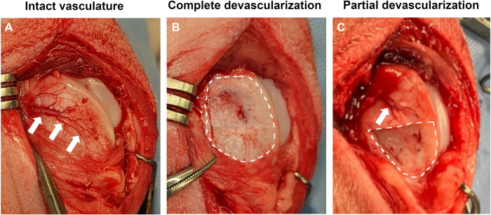
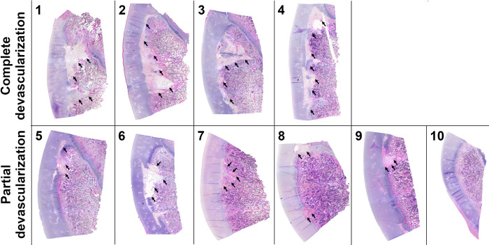
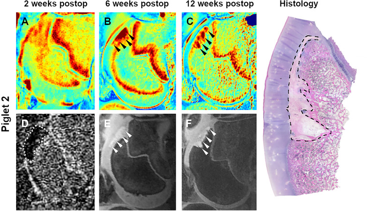
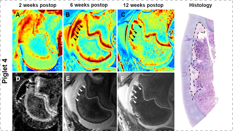
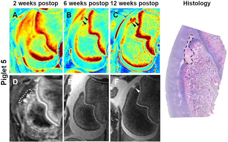
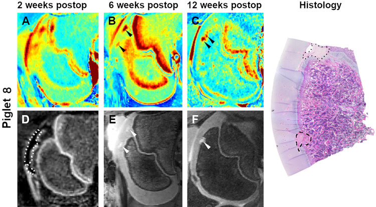
Ostechondritis dissecans (OCD) is an orthopaedic disease characterized by formation of osteochondral defects in developing joints. Epiphyseal cartilage necrosis (osteochondrosis [OC]) caused by focal failure of vascular supply is the known precursor lesion of OCD, but it remains to be established how the severity of vascular failure drives lesion healing or progression. In the current study we have implemented a novel piglet model of induced osteochondrosis of the lateral trochlear ridge of the femur to determine the role that the extent of ischemia plays in the development and progression of OC/OCD lesions. Ten 4-week-old Yorkshire piglets underwent surgical interruption of the vascular supply to the entirety (n = 4 pigs) or the distal half (n = 6 pigs) of the lateral trochlear ridge of the femur. At 2, 6, and 12 weeks postoperatively, distal femora were evaluated by magnetic resonance imaging (MRI) to determine the fate of induced OC lesions. At 12 weeks, piglets were euthanized, and the surgical sites were examined histologically. After complete devascularization, lesion size increased between the 6- and 12-week MRI by an average of 24.8 mm2 (95% CI: [-2.2, 51.7]; p = 0.071). During the same period, lesion size decreased by an average of 7.6 mm2 (95% CI: [-24.5, 19.4]; p = 0.83) in piglets receiving partial devascularization. At 12 weeks, average ± SD lesion size was larger (p<0.001) in piglets undergoing complete (73.5 ± 17.6 mm2) vs. partial (16.5 ± 9.8 mm2) devascularization. Our study demonstrates how the degree of vascular interruption determines lesion size and likelihood of healing in a large animal model of trochlear OC.
剥脱性骨软骨炎(OCD)是一种骨科疾病,其特征是在发育中的关节中形成骨软骨缺损。由局部血供衰竭引起的骨骺软骨坏死(骨软骨病[OC])是已知的OCD前驱病变,但血供衰竭的严重程度如何驱动病变愈合或进展仍有待确定。在本研究中,我们实施了一种新型的仔猪股骨外侧滑车嵴诱导性骨软骨病模型,以确定缺血程度在OC/OCD病变发展和进展中所起的作用。十只4周龄的约克夏仔猪接受手术,阻断股骨外侧滑车嵴全部(n = 4头猪)或远端一半(n = 6头猪)的血供。术后2周、6周和12周,通过磁共振成像(MRI)评估股骨远端,以确定诱导性OC病变的转归。12周时,对仔猪实施安乐死,并对手术部位进行组织学检查。完全缺血后,病变大小在6周和12周MRI检查之间平均增加24.8 mm2(95% CI:[-2.2, 51.7];p = 0.071)。在同一时期,接受部分缺血的仔猪病变大小平均减少7.6 mm2(95% CI:[-24.5, 19.4];p = 0.83)。12周时,完全缺血(73.5 ± 17.6 mm2)与部分缺血(16.5 ± 9.8 mm2)的仔猪相比,平均±标准差病变大小更大(p<0.001)。我们的研究表明,在滑车OC的大型动物模型中,血管阻断程度如何决定病变大小和愈合可能性。