• 文献检索
  • 文档翻译
  • 深度研究
  • 学术资讯
  • Suppr Zotero 插件Zotero 插件
  • 邀请有礼
  • 套餐&价格
  • 历史记录
应用&插件
Suppr Zotero 插件Zotero 插件浏览器插件Mac 客户端Windows 客户端微信小程序
定价
高级版会员购买积分包购买API积分包
服务
文献检索文档翻译深度研究API 文档MCP 服务
关于我们
关于 Suppr公司介绍联系我们用户协议隐私条款
关注我们

Suppr 超能文献

核心技术专利:CN118964589B侵权必究
粤ICP备2023148730 号-1Suppr @ 2026

文献检索

告别复杂PubMed语法,用中文像聊天一样搜索,搜遍4000万医学文献。AI智能推荐,让科研检索更轻松。

立即免费搜索

文件翻译

保留排版,准确专业,支持PDF/Word/PPT等文件格式,支持 12+语言互译。

免费翻译文档

深度研究

AI帮你快速写综述,25分钟生成高质量综述,智能提取关键信息,辅助科研写作。

立即免费体验

单细胞分辨率下发育中的人类新皮质的分子与细胞动力学

Molecular and cellular dynamics of the developing human neocortex at single-cell resolution.

作者信息

Wang Li, Wang Cheng, Moriano Juan A, Chen Songcang, Zuo Guolong, Cebrián-Silla Arantxa, Zhang Shaobo, Mukhtar Tanzila, Wang Shaohui, Song Mengyi, de Oliveira Lilian Gomes, Bi Qiuli, Augustin Jonathan J, Ge Xinxin, Paredes Mercedes F, Huang Eric J, Alvarez-Buylla Arturo, Duan Xin, Li Jingjing, Kriegstein Arnold R

机构信息

The Eli and Edythe Broad Center of Regeneration Medicine and Stem Cell Research, University of California San Francisco; San Francisco, CA 94143, USA.

Department of Neurology, University of California San Francisco; San Francisco, CA 94143, USA.

出版信息

bioRxiv. 2024 Aug 4:2024.01.16.575956. doi: 10.1101/2024.01.16.575956.

DOI:10.1101/2024.01.16.575956
PMID:39131371
原文链接:https://pmc.ncbi.nlm.nih.gov/articles/PMC11312442/
Abstract

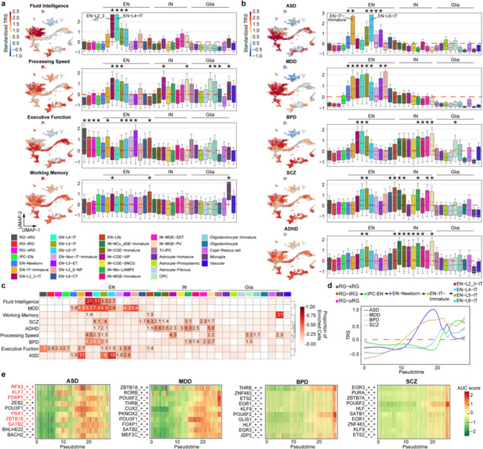
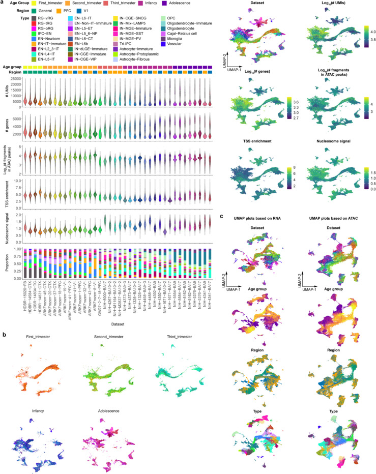
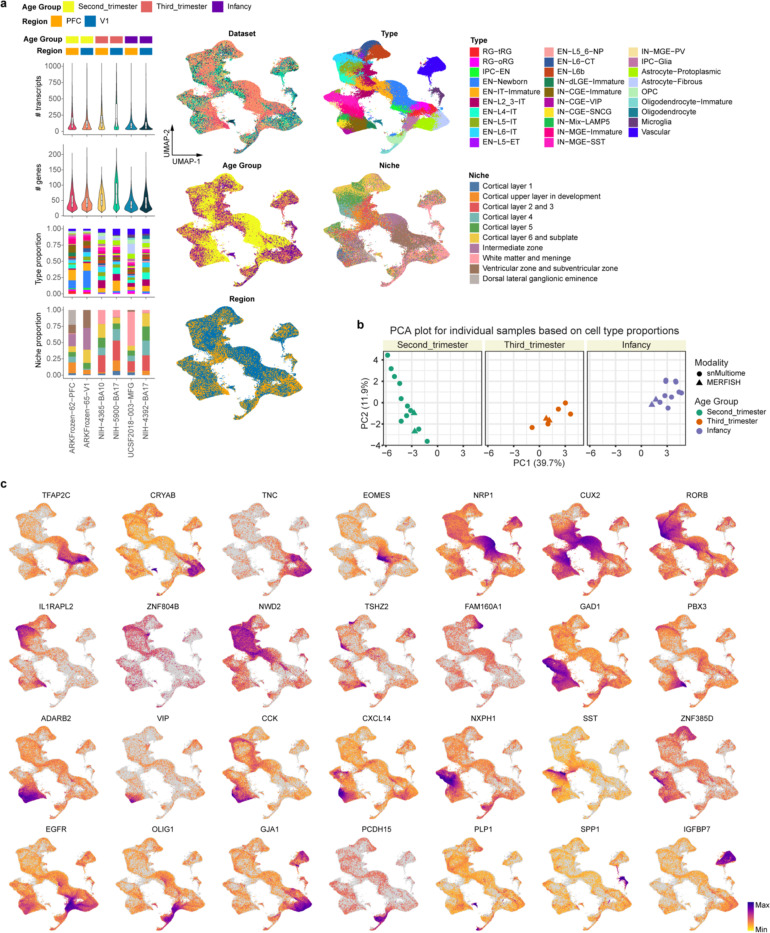
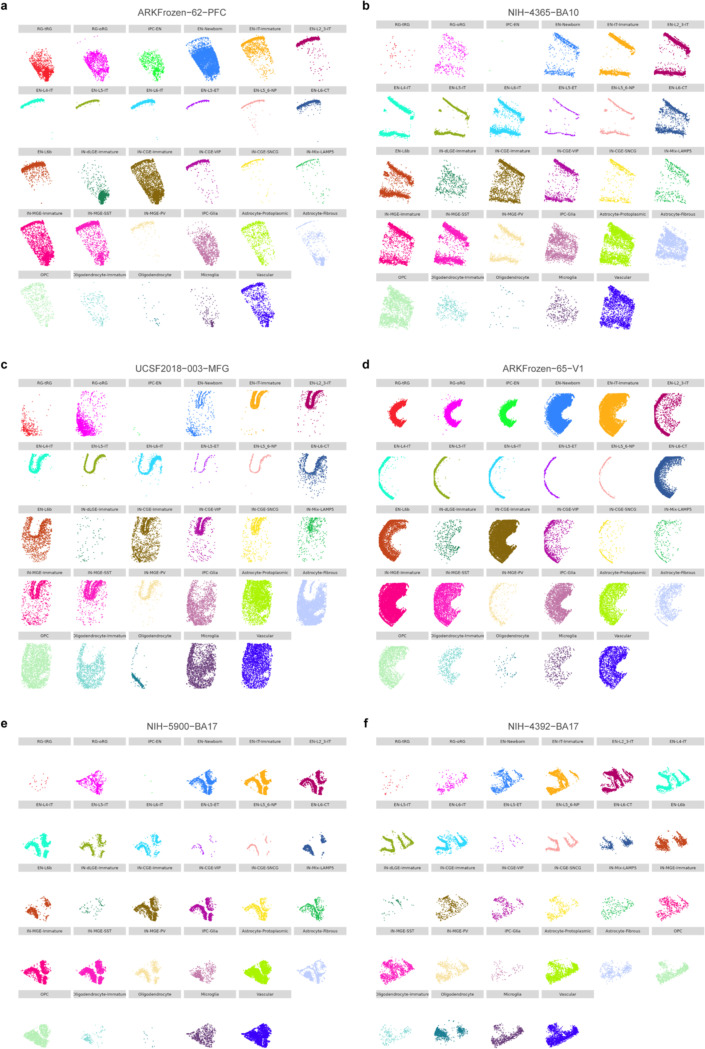
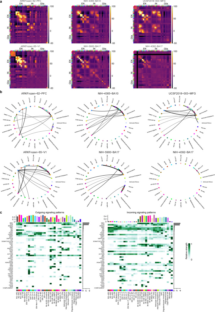
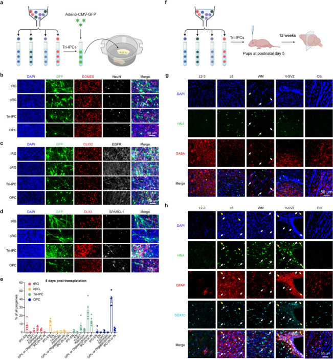
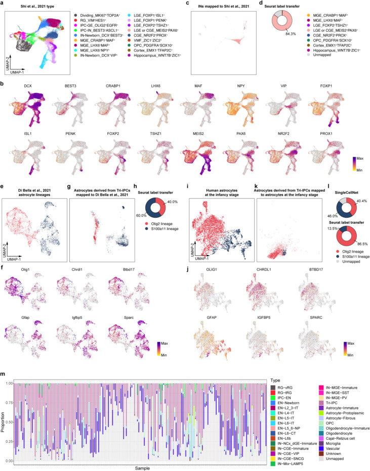
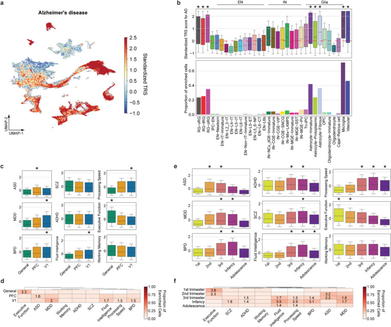
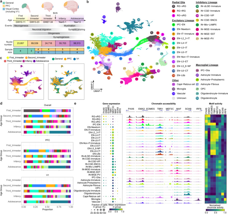
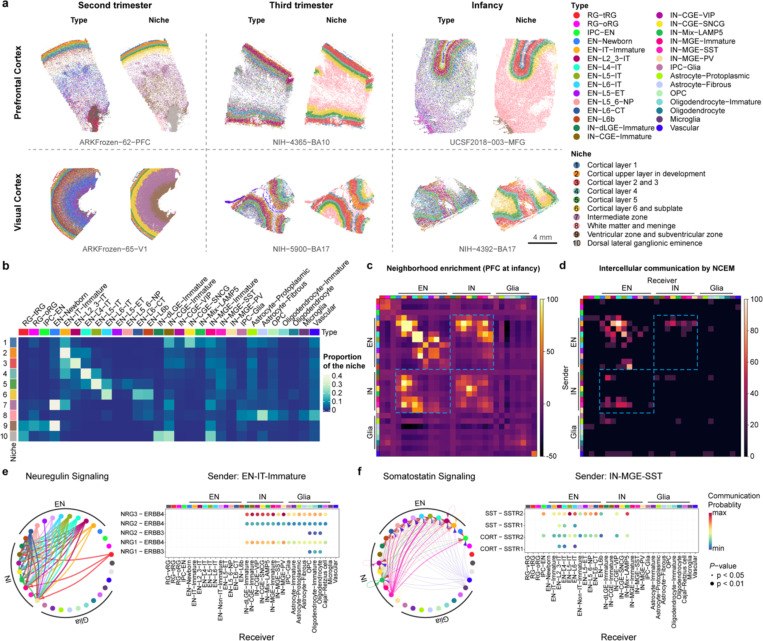
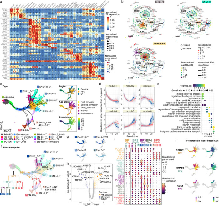
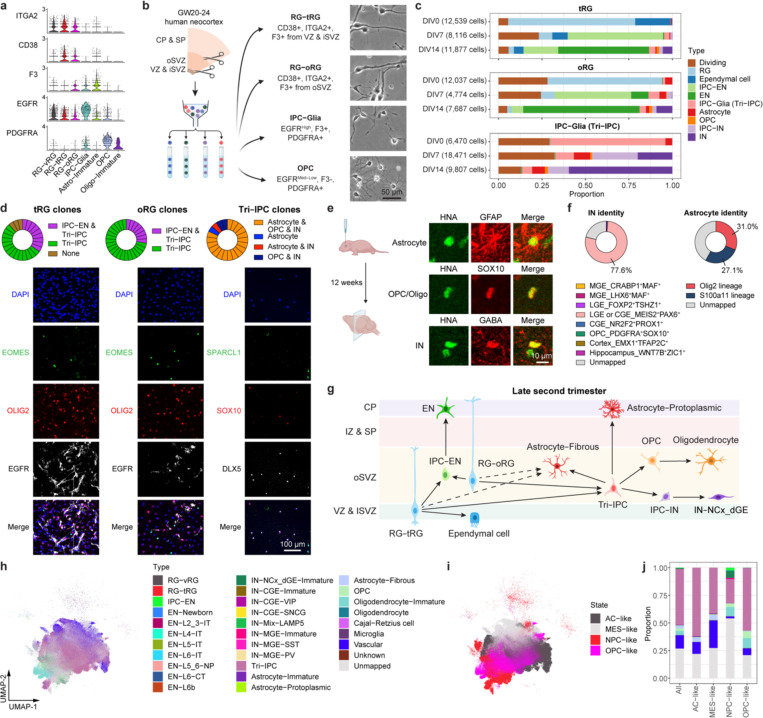
The development of the human neocortex is a highly dynamic process and involves complex cellular trajectories controlled by cell-type-specific gene regulation. Here, we collected paired single-nucleus chromatin accessibility and transcriptome data from 38 human neocortical samples encompassing both the prefrontal cortex and primary visual cortex. These samples span five main developmental stages, ranging from the first trimester to adolescence. In parallel, we performed spatial transcriptomic analysis on a subset of the samples to illustrate spatial organization and intercellular communication. This atlas enables us to catalog cell type-, age-, and area-specific gene regulatory networks underlying neural differentiation. Moreover, combining single-cell profiling, progenitor purification, and lineage-tracing experiments, we have untangled the complex lineage relationships among progenitor subtypes during the transition from neurogenesis to gliogenesis in the human neocortex. We identified a tripotential intermediate progenitor subtype, termed Tri-IPC, responsible for the local production of GABAergic neurons, oligodendrocyte precursor cells, and astrocytes. Remarkably, most glioblastoma cells resemble Tri-IPCs at the transcriptomic level, suggesting that cancer cells hijack developmental processes to enhance growth and heterogeneity. Furthermore, by integrating our atlas data with large-scale GWAS data, we created a disease-risk map highlighting enriched ASD risk in second-trimester intratelencephalic projection neurons. Our study sheds light on the gene regulatory landscape and cellular dynamics of the developing human neocortex.

摘要

人类新皮质的发育是一个高度动态的过程,涉及由细胞类型特异性基因调控控制的复杂细胞轨迹。在这里,我们从38个人类新皮质样本中收集了配对的单核染色质可及性和转录组数据,这些样本涵盖前额叶皮质和初级视觉皮质。这些样本跨越五个主要发育阶段,从妊娠早期到青春期。同时,我们对一部分样本进行了空间转录组分析,以阐明空间组织和细胞间通讯。这个图谱使我们能够编目神经分化背后的细胞类型、年龄和区域特异性基因调控网络。此外,结合单细胞分析、祖细胞纯化和谱系追踪实验,我们理清了人类新皮质从神经发生向胶质发生转变过程中祖细胞亚型之间复杂的谱系关系。我们鉴定出一种三能中间祖细胞亚型,称为Tri-IPC,它负责局部产生GABA能神经元、少突胶质细胞前体细胞和星形胶质细胞。值得注意的是,大多数胶质母细胞瘤细胞在转录组水平上类似于Tri-IPC,这表明癌细胞劫持发育过程以增强生长和异质性。此外,通过将我们的图谱数据与大规模全基因组关联研究(GWAS)数据相结合,我们创建了一个疾病风险图,突出了妊娠中期脑内投射神经元中丰富的自闭症谱系障碍(ASD)风险。我们的研究揭示了发育中的人类新皮质的基因调控格局和细胞动态。

https://cdn.ncbi.nlm.nih.gov/pmc/blobs/71ca/11312442/115fc8683627/nihpp-2024.01.16.575956v2-f0005.jpg
https://cdn.ncbi.nlm.nih.gov/pmc/blobs/71ca/11312442/f43aeff6e306/nihpp-2024.01.16.575956v2-f0006.jpg
https://cdn.ncbi.nlm.nih.gov/pmc/blobs/71ca/11312442/58b61ce5b112/nihpp-2024.01.16.575956v2-f0007.jpg
https://cdn.ncbi.nlm.nih.gov/pmc/blobs/71ca/11312442/849d0718b677/nihpp-2024.01.16.575956v2-f0008.jpg
https://cdn.ncbi.nlm.nih.gov/pmc/blobs/71ca/11312442/65d6153c1ab3/nihpp-2024.01.16.575956v2-f0009.jpg
https://cdn.ncbi.nlm.nih.gov/pmc/blobs/71ca/11312442/ad13116fda24/nihpp-2024.01.16.575956v2-f0010.jpg
https://cdn.ncbi.nlm.nih.gov/pmc/blobs/71ca/11312442/5c5e770c95c8/nihpp-2024.01.16.575956v2-f0011.jpg
https://cdn.ncbi.nlm.nih.gov/pmc/blobs/71ca/11312442/a7e7740e30b2/nihpp-2024.01.16.575956v2-f0012.jpg
https://cdn.ncbi.nlm.nih.gov/pmc/blobs/71ca/11312442/913faa01fcb4/nihpp-2024.01.16.575956v2-f0013.jpg
https://cdn.ncbi.nlm.nih.gov/pmc/blobs/71ca/11312442/b8b109c92242/nihpp-2024.01.16.575956v2-f0014.jpg
https://cdn.ncbi.nlm.nih.gov/pmc/blobs/71ca/11312442/7b104ee8074b/nihpp-2024.01.16.575956v2-f0015.jpg
https://cdn.ncbi.nlm.nih.gov/pmc/blobs/71ca/11312442/e4cc5ea90238/nihpp-2024.01.16.575956v2-f0016.jpg
https://cdn.ncbi.nlm.nih.gov/pmc/blobs/71ca/11312442/b8a558cebf19/nihpp-2024.01.16.575956v2-f0017.jpg
https://cdn.ncbi.nlm.nih.gov/pmc/blobs/71ca/11312442/da3e2c27c81a/nihpp-2024.01.16.575956v2-f0018.jpg
https://cdn.ncbi.nlm.nih.gov/pmc/blobs/71ca/11312442/20190c948a9d/nihpp-2024.01.16.575956v2-f0019.jpg
https://cdn.ncbi.nlm.nih.gov/pmc/blobs/71ca/11312442/18450e929a5b/nihpp-2024.01.16.575956v2-f0020.jpg
https://cdn.ncbi.nlm.nih.gov/pmc/blobs/71ca/11312442/f5323990f3fa/nihpp-2024.01.16.575956v2-f0021.jpg
https://cdn.ncbi.nlm.nih.gov/pmc/blobs/71ca/11312442/d50575616d81/nihpp-2024.01.16.575956v2-f0022.jpg
https://cdn.ncbi.nlm.nih.gov/pmc/blobs/71ca/11312442/3b143e47132d/nihpp-2024.01.16.575956v2-f0023.jpg
https://cdn.ncbi.nlm.nih.gov/pmc/blobs/71ca/11312442/98587679302f/nihpp-2024.01.16.575956v2-f0001.jpg
https://cdn.ncbi.nlm.nih.gov/pmc/blobs/71ca/11312442/fe33b317e00d/nihpp-2024.01.16.575956v2-f0002.jpg
https://cdn.ncbi.nlm.nih.gov/pmc/blobs/71ca/11312442/7b0dfa5fe15d/nihpp-2024.01.16.575956v2-f0003.jpg
https://cdn.ncbi.nlm.nih.gov/pmc/blobs/71ca/11312442/5ba1149f1747/nihpp-2024.01.16.575956v2-f0004.jpg
https://cdn.ncbi.nlm.nih.gov/pmc/blobs/71ca/11312442/115fc8683627/nihpp-2024.01.16.575956v2-f0005.jpg

相似文献

1
Molecular and cellular dynamics of the developing human neocortex at single-cell resolution.单细胞分辨率下发育中的人类新皮质的分子与细胞动力学
bioRxiv. 2024 Aug 4:2024.01.16.575956. doi: 10.1101/2024.01.16.575956.
2
Molecular and cellular dynamics of the developing human neocortex.发育中的人类新皮层的分子与细胞动力学
Nature. 2025 Jan 8. doi: 10.1038/s41586-024-08351-7.
3
Distinct progenitor behavior underlying neocortical gliogenesis related to tumorigenesis.与肿瘤发生相关的新皮质神经胶质生成的独特祖细胞行为。
Cell Rep. 2021 Mar 16;34(11):108853. doi: 10.1016/j.celrep.2021.108853.
4
Developmental isoform diversity in the human neocortex informs neuropsychiatric risk mechanisms.人类新皮层中的发育异构体多样性揭示神经精神疾病风险机制。
bioRxiv. 2023 Oct 11:2023.03.25.534016. doi: 10.1101/2023.03.25.534016.
5
The characteristics of excitatory lineage differentiation and the developmental conservation in Reeler neocortex.Reeler 新皮层中兴奋性谱系分化的特征及发育保守性。
Cell Prolif. 2024 May;57(5):e13587. doi: 10.1111/cpr.13587. Epub 2023 Dec 12.
6
Temporal transcriptomic dynamics in developing macaque neocortex.灵长类动物新皮层发育过程中的时间转录组动态。
Elife. 2024 Feb 28;12:RP90325. doi: 10.7554/eLife.90325.
7
Cortical Neural Stem Cell Lineage Progression Is Regulated by Extrinsic Signaling Molecule Sonic Hedgehog.皮质神经干细胞谱系的进展受外源性信号分子 Sonic Hedgehog 的调控。
Cell Rep. 2020 Mar 31;30(13):4490-4504.e4. doi: 10.1016/j.celrep.2020.03.027.
8
A Single-Cell Transcriptomic Atlas of Human Neocortical Development during Mid-gestation.人类皮质中期发育的单细胞转录组图谱。
Neuron. 2019 Sep 4;103(5):785-801.e8. doi: 10.1016/j.neuron.2019.06.011. Epub 2019 Jul 11.
9
A multi-omic single-cell landscape of cellular diversification in the developing human cerebral cortex.发育中的人类大脑皮层细胞多样化的多组学单细胞图谱。
Comput Struct Biotechnol J. 2024 May 17;23:2173-2189. doi: 10.1016/j.csbj.2024.05.019. eCollection 2024 Dec.
10
Regulation of temporal properties of neural stem cells and transition timing of neurogenesis and gliogenesis during mammalian neocortical development.调控哺乳动物新皮质发育过程中神经干细胞的时间特性和神经发生与神经胶质发生的转变时机。
Semin Cell Dev Biol. 2019 Nov;95:4-11. doi: 10.1016/j.semcdb.2019.01.007. Epub 2019 Jan 17.

引用本文的文献

1
Anatomical and molecular development of the human primary visual cortex.人类初级视觉皮层的解剖学和分子发育
Front Cell Neurosci. 2024 Sep 30;18:1427515. doi: 10.3389/fncel.2024.1427515. eCollection 2024.

本文引用的文献

1
Multiomic profiling of transcription factor binding and function in human brain.人类大脑中转录因子结合和功能的多组学分析。
Nat Neurosci. 2024 Jul;27(7):1387-1399. doi: 10.1038/s41593-024-01658-8. Epub 2024 Jun 3.
2
Chromatin accessibility during human first-trimester neurodevelopment.人类孕早期神经发育过程中的染色质可及性。
Nature. 2024 May 1. doi: 10.1038/s41586-024-07234-1.
3
Spatiotemporal transcriptome atlas reveals the regional specification of the developing human brain.时空转录组图谱揭示了人类大脑发育的区域特化。
Cell. 2023 Dec 21;186(26):5892-5909.e22. doi: 10.1016/j.cell.2023.11.016. Epub 2023 Dec 12.
4
Transcriptomic cytoarchitecture reveals principles of human neocortex organization.转录组细胞构筑揭示了人类新皮层组织的原则。
Science. 2023 Oct 13;382(6667):eadf6812. doi: 10.1126/science.adf6812.
5
Comprehensive cell atlas of the first-trimester developing human brain.人类大脑第一孕期发育的全面细胞图谱。
Science. 2023 Oct 13;382(6667):eadf1226. doi: 10.1126/science.adf1226.
6
Single-cell analysis of prenatal and postnatal human cortical development.单细胞分析人类产前和产后皮质发育。
Science. 2023 Oct 13;382(6667):eadf0834. doi: 10.1126/science.adf0834.
7
A comparative atlas of single-cell chromatin accessibility in the human brain.人类大脑单细胞染色质可及性比较图谱。
Science. 2023 Oct 13;382(6667):eadf7044. doi: 10.1126/science.adf7044.
8
A marmoset brain cell census reveals regional specialization of cellular identities.狨猴脑细胞普查揭示了细胞特征的区域专业化。
Sci Adv. 2023 Oct 13;9(41):eadk3986. doi: 10.1126/sciadv.adk3986. Epub 2023 Oct 12.
9
Multi-omic profiling of the developing human cerebral cortex at the single-cell level.单细胞水平上人类大脑皮质发育的多组学分析。
Sci Adv. 2023 Oct 13;9(41):eadg3754. doi: 10.1126/sciadv.adg3754. Epub 2023 Oct 12.
10
LIF signaling regulates outer radial glial to interneuron fate during human cortical development.LIF 信号在人皮质发育过程中调节外放射状胶质向中间神经元的命运。
Cell Stem Cell. 2023 Oct 5;30(10):1382-1391.e5. doi: 10.1016/j.stem.2023.08.009. Epub 2023 Sep 5.