• 文献检索
  • 文档翻译
  • 深度研究
  • 学术资讯
  • Suppr Zotero 插件Zotero 插件
  • 邀请有礼
  • 套餐&价格
  • 历史记录
应用&插件
Suppr Zotero 插件Zotero 插件浏览器插件Mac 客户端Windows 客户端微信小程序
定价
高级版会员购买积分包购买API积分包
服务
文献检索文档翻译深度研究API 文档MCP 服务
关于我们
关于 Suppr公司介绍联系我们用户协议隐私条款
关注我们

Suppr 超能文献

核心技术专利:CN118964589B侵权必究
粤ICP备2023148730 号-1Suppr @ 2026

文献检索

告别复杂PubMed语法,用中文像聊天一样搜索,搜遍4000万医学文献。AI智能推荐,让科研检索更轻松。

立即免费搜索

文件翻译

保留排版,准确专业,支持PDF/Word/PPT等文件格式,支持 12+语言互译。

免费翻译文档

深度研究

AI帮你快速写综述,25分钟生成高质量综述,智能提取关键信息,辅助科研写作。

立即免费体验

斑马鱼颅缝建立过程中的细胞转变。

Cellular transitions during cranial suture establishment in zebrafish.

机构信息

Department of Molecular, Cell and Developmental Biology, University of California, Los Angeles, CA, 90095, USA.

Eli and Edythe Broad Center for Regenerative Medicine, Department of Stem Cell Biology and Regenerative Medicine, Keck School of Medicine, University of Southern California, Los Angeles, CA, 90033, USA.

出版信息

Nat Commun. 2024 Aug 13;15(1):6948. doi: 10.1038/s41467-024-50780-5.

DOI:10.1038/s41467-024-50780-5
PMID:39138165
原文链接:https://pmc.ncbi.nlm.nih.gov/articles/PMC11322166/
Abstract

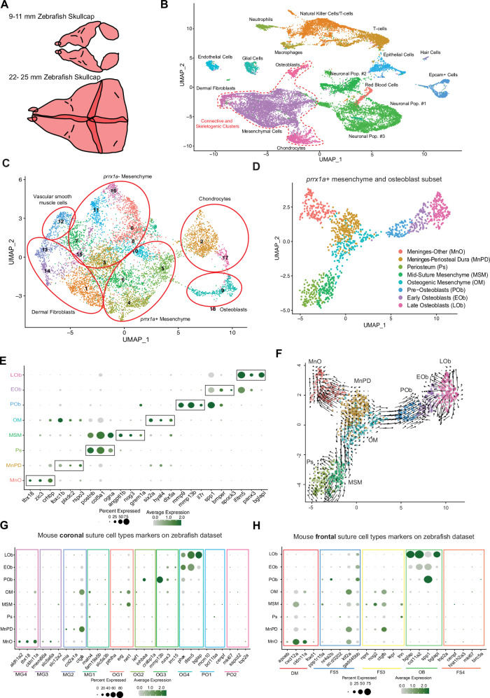
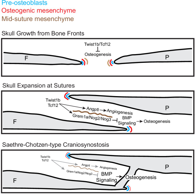
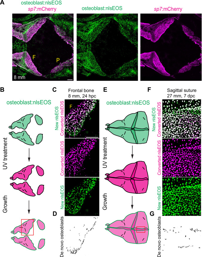
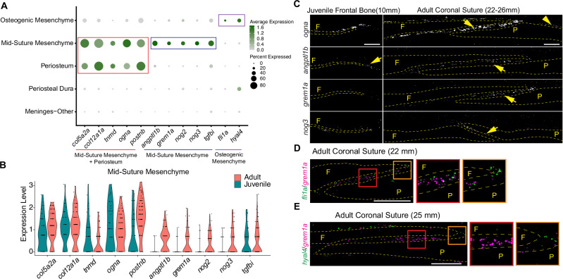
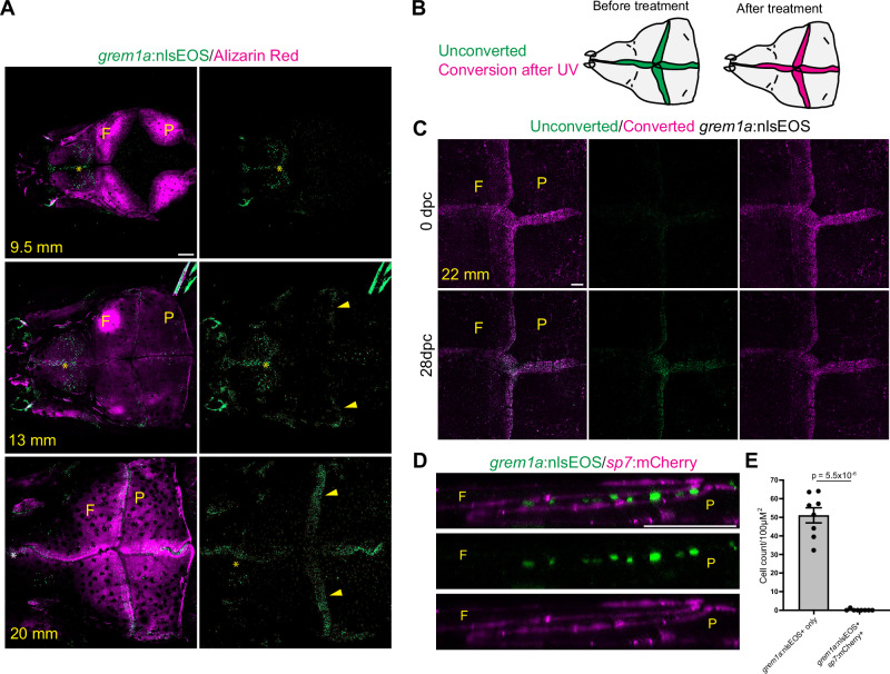
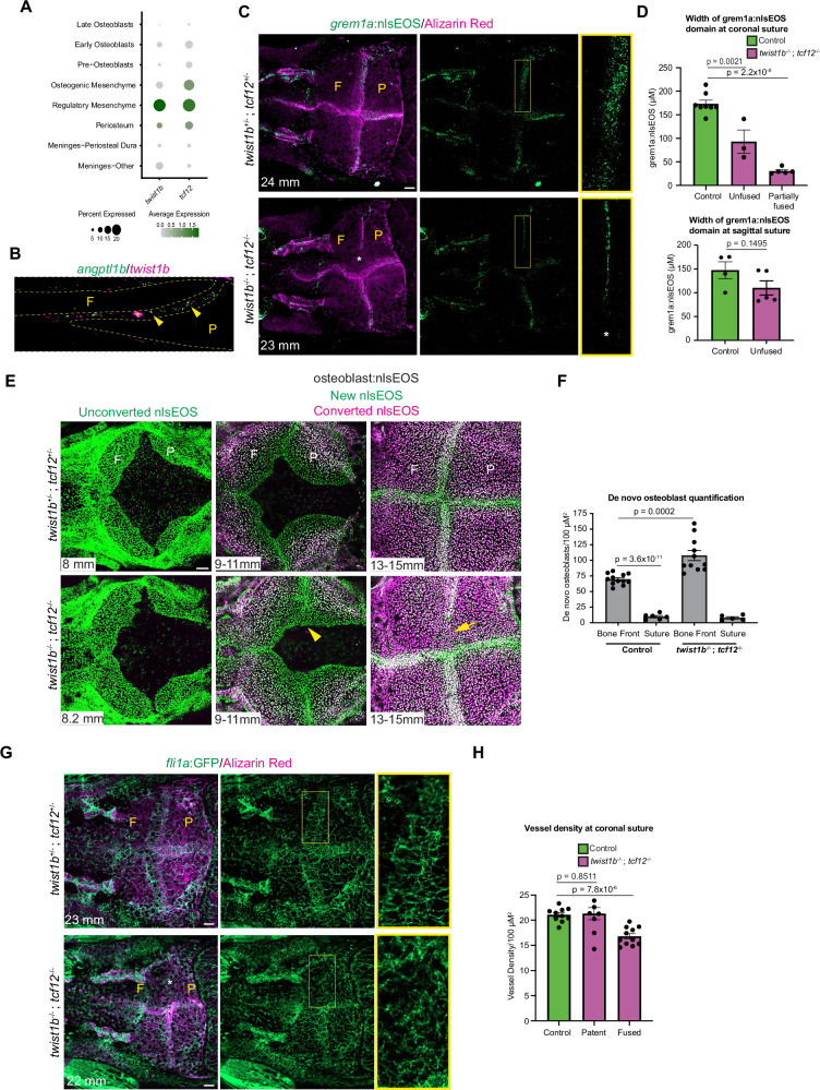
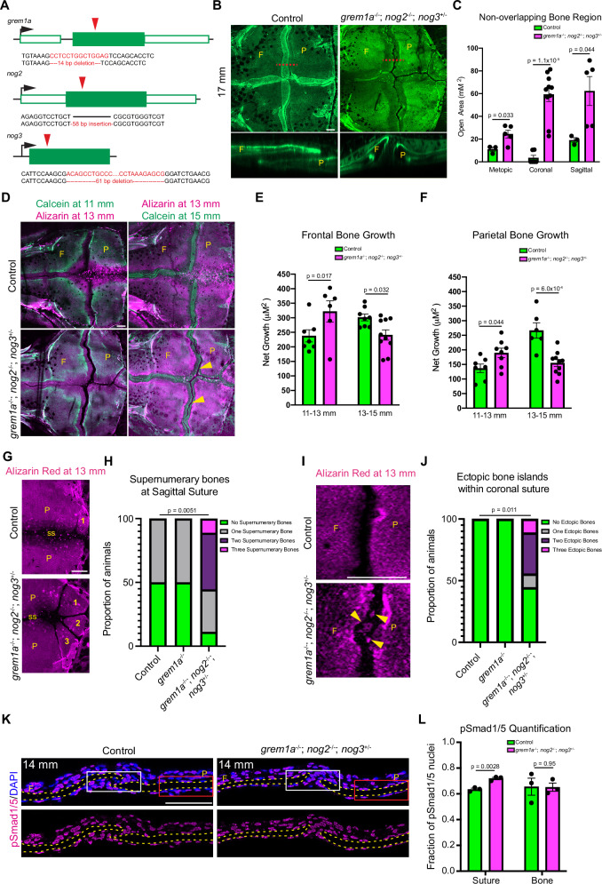
Cranial sutures separate neighboring skull bones and are sites of bone growth. A key question is how osteogenic activity is controlled to promote bone growth while preventing aberrant bone fusions during skull expansion. Using single-cell transcriptomics, lineage tracing, and mutant analysis in zebrafish, we uncover key developmental transitions regulating bone formation at sutures during skull expansion. In particular, we identify a subpopulation of mesenchyme cells in the mid-suture region that upregulate a suite of genes including BMP antagonists (e.g. grem1a) and pro-angiogenic factors. Lineage tracing with grem1a:nlsEOS reveals that this mid-suture subpopulation is largely non-osteogenic. Moreover, combinatorial mutation of BMP antagonists enriched in this mid-suture subpopulation results in increased BMP signaling in the suture, misregulated bone formation, and abnormal suture morphology. These data reveal establishment of a non-osteogenic mesenchyme population in the mid-suture region that restricts bone formation through local BMP antagonism, thus ensuring proper suture morphology.

摘要

颅缝分隔相邻的颅骨,并为骨骼生长提供场所。一个关键问题是如何控制成骨活性,以促进骨骼生长,同时防止颅骨扩张过程中的异常骨融合。通过在斑马鱼中使用单细胞转录组学、谱系追踪和突变分析,我们揭示了调节颅骨扩张过程中颅缝骨形成的关键发育转变。特别是,我们在中缝区域鉴定出一群间充质细胞,这些细胞上调了包括 BMP 拮抗剂(例如 grem1a)和促血管生成因子在内的一系列基因。利用 grem1a:nlsEOS 进行的谱系追踪表明,这个中缝亚群在很大程度上是非成骨的。此外,在这个中缝亚群中富集的 BMP 拮抗剂的组合突变导致在颅缝中 BMP 信号的增加、骨形成的失调和异常的颅缝形态。这些数据揭示了在中缝区域建立了一个非成骨的间充质细胞群体,通过局部 BMP 拮抗作用限制骨形成,从而确保适当的颅缝形态。

https://cdn.ncbi.nlm.nih.gov/pmc/blobs/e98a/11322166/a20e7c8f27ef/41467_2024_50780_Fig9_HTML.jpg
https://cdn.ncbi.nlm.nih.gov/pmc/blobs/e98a/11322166/b53524a0a958/41467_2024_50780_Fig1_HTML.jpg
https://cdn.ncbi.nlm.nih.gov/pmc/blobs/e98a/11322166/2452ba64ebaf/41467_2024_50780_Fig2_HTML.jpg
https://cdn.ncbi.nlm.nih.gov/pmc/blobs/e98a/11322166/be56248d34fc/41467_2024_50780_Fig3_HTML.jpg
https://cdn.ncbi.nlm.nih.gov/pmc/blobs/e98a/11322166/e7f240ecddb5/41467_2024_50780_Fig4_HTML.jpg
https://cdn.ncbi.nlm.nih.gov/pmc/blobs/e98a/11322166/d1093f88465b/41467_2024_50780_Fig5_HTML.jpg
https://cdn.ncbi.nlm.nih.gov/pmc/blobs/e98a/11322166/aa02a02b02be/41467_2024_50780_Fig6_HTML.jpg
https://cdn.ncbi.nlm.nih.gov/pmc/blobs/e98a/11322166/4942e3eab17d/41467_2024_50780_Fig7_HTML.jpg
https://cdn.ncbi.nlm.nih.gov/pmc/blobs/e98a/11322166/a4a1ebeb98b8/41467_2024_50780_Fig8_HTML.jpg
https://cdn.ncbi.nlm.nih.gov/pmc/blobs/e98a/11322166/a20e7c8f27ef/41467_2024_50780_Fig9_HTML.jpg

相似文献

1
Cellular transitions during cranial suture establishment in zebrafish.斑马鱼颅缝建立过程中的细胞转变。
Nat Commun. 2024 Aug 13;15(1):6948. doi: 10.1038/s41467-024-50780-5.
2
The BMP ligand Gdf6 prevents differentiation of coronal suture mesenchyme in early cranial development.骨形态发生蛋白配体 Gdf6 可防止颅早期发育中冠状缝间充质的分化。
PLoS One. 2012;7(5):e36789. doi: 10.1371/journal.pone.0036789. Epub 2012 May 31.
3
FGF-, BMP- and Shh-mediated signalling pathways in the regulation of cranial suture morphogenesis and calvarial bone development.成纤维细胞生长因子、骨形态发生蛋白和音猬因子介导的信号通路在颅缝形态发生和颅骨发育调控中的作用
Development. 1998 Apr;125(7):1241-51. doi: 10.1242/dev.125.7.1241.
4
Apoptosis in murine calvarial bone and suture development.小鼠颅骨和缝线发育中的细胞凋亡
Eur J Oral Sci. 1999 Aug;107(4):265-75. doi: 10.1046/j.0909-8836.1999.eos107406.x.
5
Osterix/Sp7 limits cranial bone initiation sites and is required for formation of sutures.成骨特异性转录因子/Sp7限制颅骨起始位点,是缝线形成所必需的。
Dev Biol. 2016 May 15;413(2):160-72. doi: 10.1016/j.ydbio.2016.03.011. Epub 2016 Mar 16.
6
The developing mouse coronal suture at single-cell resolution.单细胞分辨率下发育中的小鼠冠状缝。
Nat Commun. 2021 Aug 10;12(1):4797. doi: 10.1038/s41467-021-24917-9.
7
The Morphogenesis of Cranial Sutures in Zebrafish.斑马鱼颅骨缝的形态发生
PLoS One. 2016 Nov 9;11(11):e0165775. doi: 10.1371/journal.pone.0165775. eCollection 2016.
8
Regulation of bone morphogenetic protein signalling and cranial osteogenesis by Gpc1 and Gpc3.Gpc1 和 Gpc3 对骨形态发生蛋白信号转导和颅骨发生的调节作用。
Bone. 2013 Aug;55(2):367-76. doi: 10.1016/j.bone.2013.04.013. Epub 2013 Apr 25.
9
Single-cell analysis identifies a key role for Hhip in murine coronal suture development.单细胞分析鉴定出 Hhip 在小鼠冠状缝发育中的关键作用。
Nat Commun. 2021 Dec 8;12(1):7132. doi: 10.1038/s41467-021-27402-5.
10
The Development of the Calvarial Bones and Sutures and the Pathophysiology of Craniosynostosis.颅骨及颅缝的发育与颅缝早闭的病理生理学
Curr Top Dev Biol. 2015;115:131-56. doi: 10.1016/bs.ctdb.2015.07.004. Epub 2015 Oct 1.

本文引用的文献

1
Skull progenitor cell-driven meningeal lymphatic restoration improves neurocognitive functions in craniosynostosis.颅骨祖细胞驱动的脑膜淋巴管修复改善颅缝早闭的神经认知功能。
Cell Stem Cell. 2023 Nov 2;30(11):1472-1485.e7. doi: 10.1016/j.stem.2023.09.012. Epub 2023 Oct 19.
2
A multi-stem cell basis for craniosynostosis and calvarial mineralization.颅缝早闭和颅骨矿化的多干细胞基础。
Nature. 2023 Sep;621(7980):804-812. doi: 10.1038/s41586-023-06526-2. Epub 2023 Sep 20.
3
Generation and application of endogenously floxed alleles for cell-specific knockout in zebrafish.
内源性打靶等位基因的产生及其在斑马鱼细胞特异性敲除中的应用。
Dev Cell. 2023 Nov 20;58(22):2614-2626.e7. doi: 10.1016/j.devcel.2023.07.022. Epub 2023 Aug 25.
4
Single-cell transcriptome profiling reveals diverse immune cell populations and their responses to viral infection in the spleen of zebrafish.单细胞转录组谱分析揭示了斑马鱼脾脏中多种免疫细胞群体及其对病毒感染的反应。
FASEB J. 2023 Jun;37(6):e22951. doi: 10.1096/fj.202201505RRRR.
5
Cre/ regulated conditional rescue and inactivation with zebrafish UFlip alleles generated by CRISPR-Cas9 targeted integration.通过 CRISPR-Cas9 靶向整合产生的 zebrafish UFlip 等位基因的 Cre/regulated 条件性拯救和失活。
Elife. 2022 Jun 17;11:e71478. doi: 10.7554/eLife.71478.
6
Single-cell transcriptome reveals insights into the development and function of the zebrafish ovary.单细胞转录组揭示了斑马鱼卵巢发育和功能的见解。
Elife. 2022 May 19;11:e76014. doi: 10.7554/eLife.76014.
7
Lifelong single-cell profiling of cranial neural crest diversification in zebrafish.斑马鱼颅神经嵴多样化的终生单细胞分析。
Nat Commun. 2022 Jan 10;13(1):13. doi: 10.1038/s41467-021-27594-w.
8
Embryonic requirements for Tcf12 in the development of the mouse coronal suture.胚胎中 Tcf12 对小鼠冠状缝发育的需求。
Development. 2022 Jan 1;149(1). doi: 10.1242/dev.199575. Epub 2022 Jan 4.
9
The developing mouse coronal suture at single-cell resolution.单细胞分辨率下发育中的小鼠冠状缝。
Nat Commun. 2021 Aug 10;12(1):4797. doi: 10.1038/s41467-021-24917-9.
10
Skeletal stem and progenitor cells maintain cranial suture patency and prevent craniosynostosis.骨骼干/祖细胞维持颅缝开放,防止颅缝早闭。
Nat Commun. 2021 Jul 30;12(1):4640. doi: 10.1038/s41467-021-24801-6.