Breden Sebastian, Hinterwimmer Florian, Consalvo Sarah, Neumann Jan, Knebel Carolin, von Eisenhart-Rothe Rüdiger, Burgkart Rainer H, Lenze Ulrich
Department of Orthopedics and Sports Orthopedics, Klinikum rechts der Isar, Technical University of Munich, 81675 Munich, Germany.
Institute for AI and Informatics in Medicine, Technical University of Munich, 81675 Munich, Germany.
J Clin Med. 2023 Sep 14;12(18):5960. doi: 10.3390/jcm12185960.
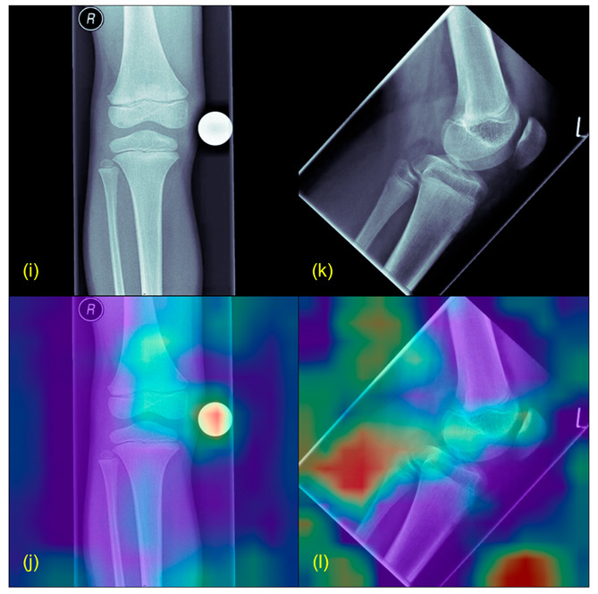
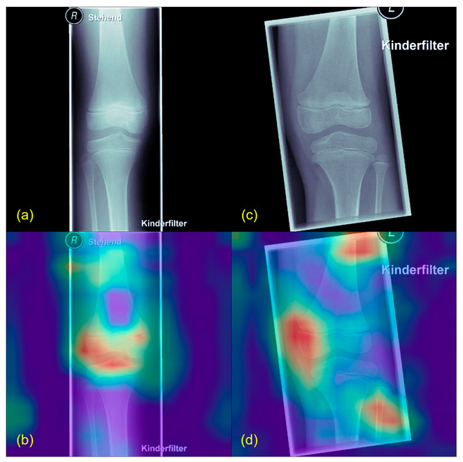
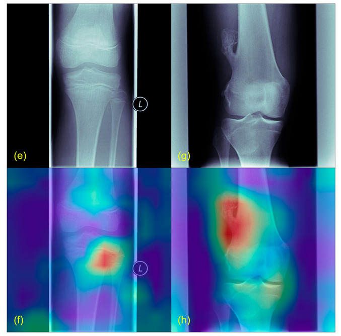
Even though tumors in children are rare, they cause the second most deaths under the age of 18 years. More often than in other age groups, underage patients suffer from malignancies of the bones, and these mostly occur in the area around the knee. One problem in the treatment is the early detection of bone tumors, especially on X-rays. The rarity and non-specific clinical symptoms further prolong the time to diagnosis. Nevertheless, an early diagnosis is crucial and can facilitate the treatment and therefore improve the prognosis of affected children. A new approach to evaluating X-ray images using artificial intelligence may facilitate the detection of suspicious lesions and, hence, accelerate the referral to a specialized center. We implemented a Vision Transformer model for image classification of healthy and pathological X-rays. To tackle the limited amount of data, we used a pretrained model and implemented extensive data augmentation. Discrete parameters were described by incidence and percentage ratio and continuous parameters by median, standard deviation and variance. For the evaluation of the model accuracy, sensitivity and specificity were computed. The two-entity classification of the healthy control group and the pathological group resulted in a cross-validated accuracy of 89.1%, a sensitivity of 82.2% and a specificity of 93.2% for test groups. Grad-CAMs were created to ensure the plausibility of the predictions. The proposed approach, using state-of-the-art deep learning methodology to detect bone tumors on knee X-rays of children has achieved very good results. With further improvement of the algorithm, enlargement of the dataset and removal of potential biases, this could become a useful additional tool, especially to support general practitioners for early, accurate and specific diagnosis of bone lesions in young patients.
尽管儿童肿瘤罕见,但它们在18岁以下人群中造成的死亡人数位居第二。与其他年龄组相比,未成年患者更常患骨恶性肿瘤,且这些肿瘤大多发生在膝盖周围区域。治疗中的一个问题是骨肿瘤的早期检测,尤其是在X光片上。其罕见性和非特异性临床症状进一步延长了诊断时间。然而,早期诊断至关重要,它可以促进治疗,从而改善患病儿童的预后。一种使用人工智能评估X光图像的新方法可能有助于检测可疑病变,进而加快转诊至专业中心的速度。我们实现了一个视觉Transformer模型,用于对健康和病理X光片进行图像分类。为了解决数据量有限的问题,我们使用了一个预训练模型并实施了广泛的数据增强。离散参数用发病率和百分比表示,连续参数用中位数、标准差和方差表示。为了评估模型的准确性,计算了灵敏度和特异性。健康对照组和病理组的二分类在测试组中交叉验证的准确率为89.1%,灵敏度为82.2%,特异性为93.2%。创建了梯度加权类激活映射以确保预测的合理性。所提出的方法,即使用最先进的深度学习方法在儿童膝盖X光片上检测骨肿瘤,已经取得了非常好的结果。随着算法的进一步改进、数据集的扩大以及潜在偏差的消除,这可能会成为一个有用的辅助工具,特别是用于支持全科医生对年轻患者的骨病变进行早期、准确和特异性的诊断。